When seeking an odor, vertebrate swimming undulates left and right, naturally moving the nose perpendicular to the body motion. This lateral motion can help navigation if odor sampling can be coordinated with the movement, enabling a spatiotemporal gradient calculation along the path of the nose movement. This lateral sampling over time is called klinotaxis (“leaning navigation”) or weathervaning.
Essay 24 and essay 25 explored head-direction navigation as inspired by the fruit fly Drosophila fan-shaped body and ellipsoid body. The idea was to use head direction to translate egocentric movement into an allocentric memory of past samples, independent of the current body direction. In contrast, klinotaxis uses an egocentric system, where the lateral motion is relative to the current direction, not an independent, compass or map-like system.
Klinotaxis in Drosophila larva and C. elegans
Klinotaxis has been largely studied in the fruit fly Drosophila larva and the roundworm C. elegans. Drosophila larva have a distinct “cast” movement, where they pause and wave their heads side to side, either a single time (1-cast) or multiple times (n-cast) [Zhao et al 2017]. Larva movements break down into five major types [Gomez-Marin and Louis 2014]:
Forward
Backward
Stop
Turn
Cast
C. elegans has two major seek movements: pirouettes and weathervaning [Lockery 2011]. Pirouettes are a u-turn when the animal is moving away from the odor. Weathervaning is a side-to-side head movement that manages turning.
Both systems are temporal gradient systems, requiring measurements at different times and a memory of the older measurement [Chen X and Engert 2014]. Klinotaxis requires a basic form of memory [Karpenko et al 2020], but the comparison can be a simple ON or OFF result [Lockery 2011]. Pirouetts use a gradient parallel to body motion and reverse direction when the animal is moving away from the odor [Iino and Yoshida 2009]. Weathervaning uses a gradient perpendicular to body motion, measured with a lateral head movement [Lockery 2011].
This klinotaxis contrasts with a bilateral spatial navigation that compares two lateral sensors [Chen X and Engert 2014], such as bilateral eyes, ears, or nostrils. In Drosophila larva, odor turning is proportional to the lateral gradient more than the parallel gradient [Martinez 2014]. The odor navigation is not simply bilateral because disabling one side of O.sn (olfactory sensory neuron) only minimally impairs navigation [Gomez-Marin and Louis 2014].
As a slight digression, let’s return to the adult Drosophila navigation, because the structure can be a useful analogy for understanding vertebrate klinotaxis navigation, despite using a different allocentric system.
Adult Drosophila FSB
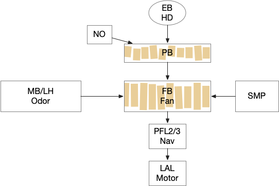
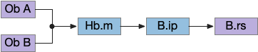
Below is a rough sketch of the Drosophila navigation circuit, focused on the fan-shaped body [Hulse et al 2021]. The ellipsoid body (EB) and protocerebral bridge (PB) calculate head direction and sort it into 18 columns. This head direction is allocentric, independent of the animal’s current direction, like a compass direction or a map. Input from odor areas like the mushroom body (MB) and lateral horn (LN) are organized into 9 rows. The fan-shaped body combines these 18 head direction columns and 9 sense data rows into a memory table.
Drosophila navigation, focusing on head direction from PB, odor data from MB and LH, and allocentric table of FB. EB ellipsoid body, FB fan-shaped body, LH lateral horn, MB mushroom body.
Motor navigation reads out from the fan-shaped-body table. These motor commands include left and right, but also include a separate u-turn command [Westeinde et al. 2022]. Although this allocentric navigation system differs from egocentric klinotaxis, its motor output includes both the left vs right from weathervaning and the u-turn from pirouette.
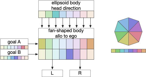
The previous essay 24 and essay 25 attempts followed this model. As the animal moves in space, the model saved the forward odor gradient according to the current head direction. By comparing stored values for other head directions, the animal would improve its heading toward the direction with the strongest odor.
The fan-shaped body then becomes a record of samples of all the older directions that the animal had measured. Output is then calculated for left (PFL3L), right (PFL3R), and u-turn (PFL2) signals. [Westeinde et al 2024]. The current head direction is represented as a sinusoidal neural pattern and combined with the stored values to produce an output.
This system was only partially successful for the essay. Although it was an improvement over no memory, because the animal was continually moving in space, the table was always obsolete. Even when the table memory times out to represent loss in accuracy as the animal moves, the rapid obsolescence made navigation difficult, particularly as the animal neared the target.
So, this essay simplifies the circuit and lowers the ambition. Instead of trying to record every direction and keeping perfect allocentric compass direction, the animal could simple save its left and right oscillation as it swims naturally.
Vertebrate Hb.m and R.ip
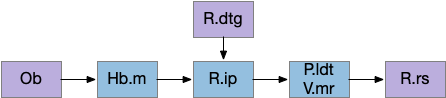
The vertebrate Hb.m (medial habenula) to R.ip (interpeduncular nucleus) is used for phototaxis [Chen X and Engert 2014], Chemotaxis [Chen WY et al 2019] and thermotaxis [Palieri et al 2024]. In a clever experiment creating a virtual light circle, Chen and Engert shows that the zebrafish phototaxis is not simply comparing light between the eyes for a spatial gradient (tropotaxis) but is a temporally-based gradient (klinotaxis), relying on a short term memory of the previous light. This phototaxis uses the Hb.m to R.ip circuit [Chen X and Engert 2014].
Head direction from R.dgt (dorsal tegmental nucleus) tiles R.ip vertically [Petrucco et al 2023], while olfactory and light input is organized horizontally [Chen WY et al 2019], [Zaupa et al 2021]. After combining the odor with the head direction and comparing with the stored values, it sends motor commands to R.rs (reticulospinal) using P.ldt (laterodorsal tegmental nucleus) and V.mr (median raphe). The vertebrate R.ip has 6 columns of head direction input from R.dtg, resembling the Drosophila fan-shaped body, but instead of 18 columns for the fan-shaped body, R.ip only has 6, three to a side [Petrucco et al 2023].
Essay 25 explored a model which used the Drosophila fan-shaped body allocentric navigation in R.ip with some limited but not overwhelming success. Instead, this essay will try a different interpretation, where R.ip is only storing side to side weathervaning of the head while swimming, instead of a full 360 degree table like Drosophila.
Vertebrate klinotaxis
As a different approach, suppose the head direction to R.ip is not an allocentric map-making coordinator as in the adult Drosophila, but a simpler egocentric weathervaning or casting coordinator, storing only the lateral gradient from head direction changes from natural swimming, or possibly deliberate larger turns like casting to gather wider lateral gradient information.
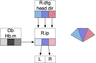
Klinotaxis simplifies the need for precise head direction. Instead of the Drosophila 18 head direction columns calibrated to the outside world, we use only three, two lateral and one central, that only require motor efference copies of left and right muscle turns. Studies from the zebrafish R.ip suggest three columns to a side, which isn’t connected to the vestibular system [Petrucco et al 2023]. To me, this suggests to me that the head direction might not be an allocentric signal that requires precise direction, but a simple egocentric lateral measurement, which doesn’t need vestibular information.
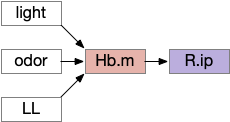
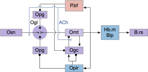
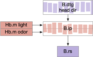
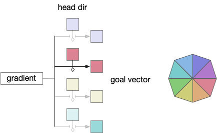
The above diagram illustrates the system. Olfactory samples arrive through Hb.mand head direction arrives from R.dtg. Like the Drosophila fan-shaped body, R.ip combines odor samples with lateral head movement into a simple memory table, and it reads out left and right motor commands. A similar system can save odor measurements parallel to body movement, using velocity instead of head direction, to trigger a u-turn when the animal is moving away from the odor.
Discussion
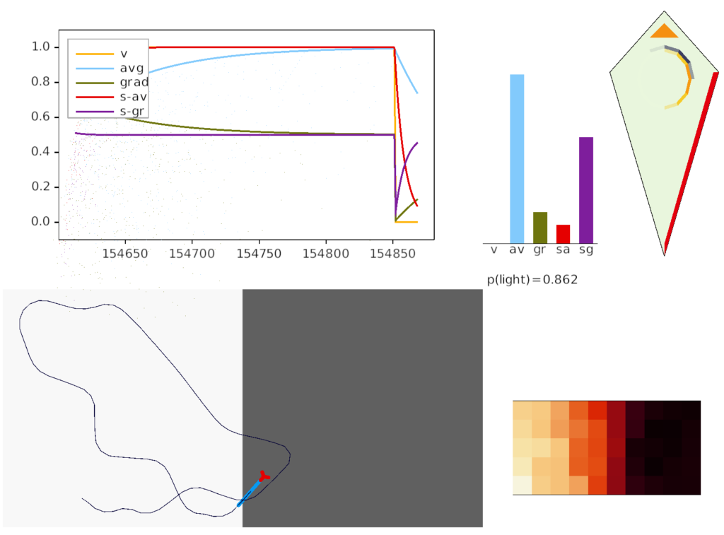
Compared to the parallel-only gradient, allocentric system of essay 25, this lateral navigation is far simpler and more effective. Even with only three bins compared to the 8 bins in essay 25, the lateral weathervaning turned out to be more effective and less brittle. If R.ip does implement a lateral klinotaxis system like this essay, it’s plausible that the 6 directions reported by [Westeinde et al 2024] are sufficient for accurate seek navigation. In contract, those 6 directions seem insufficient for an allocentric navigation compared to the Drosophila 18 directions.
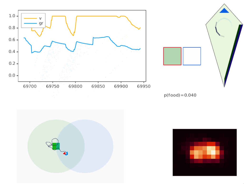
Interestingly, the pirouette also highly effective, even without lateral klinotaxis. In the simulation, when the animal moved away from the odor source, it makes a u-turn. This system served to ratchet the animal closer and closer to the target. Even when most of the movement was random, the pirouette locks in any improvement. Pirouette itself is also simple, only requiring two averages: a short average and a long average, where a short average tracks the odor across a single swim cycle and a long average uses two swim cycles. When the short average has a stronger odor value than the long average, the animal is moving toward the odor.
In both cases, the simulation used a binary OFF for the motor command instead of attempting finer precision from the gradient. This simple OFF strategy was sufficient for the simulation. A C. elegans study suggested that ON-OFF coding was energy efficient, and the worm rarely orients perfectly to the gradient [Lockery 2011].
I’m looking to improve the foraging algorithm with an idea from essay 17, which suggested that when the foraging fails, the animal should avoid the failed area. The foraging task uses an odor cue to seek food. Currently when the model gives up (times out), it disables seeking, but doesn’t actively avoid the current place, but returns to the wide-ranging roaming search.
For now, I’m still avoiding memory, but consider the alternating T-maze used in rodent behavior [Deacon and Rawlins 2006]. Mice are released at the base of the T and choose one of the directions to search for food. If the experiment repeats (by picking up the mice and restarting) mice will tend to explore the unexplored end first.
But for our foraging task, let’s use the same device for a different purpose. Instead of repeating the experiment by unnatural teleportation, consider the simpler problem of foraging with this device as an environment.
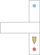
T-maze exploration. Food might be at either the red dot or the blue dot.
When rodents are foraging and reach one end, they will reverse and search the other end. Because rodents are far more advanced than the toy model, they can remember which arm of the maze they’ve already explored. But consider a simpler sub-strategy that uses RTPA (real-time place avoidance) where the animal temporarily avoids the current area or areas associated with food. By actively avoiding the already-explored area, the animal will save time by avoiding repeated searching.
A difficulty in finding the neural correlates of RTPA is the great diversity of reasons for RTPA, and circuits even in the brainstem. There are many reasons for place avoidance:
Startle: reflex escape
Escape from an imminent predator
Escape from an environment hazard (CO2, temperature)
Avoiding innate cues (predator odors)
Avoiding learned cues (CPA conditioned place avoidance)
Search optimization: avoiding already searched areas
Because this topic is large and the number of circuits is also large, I’ll start with a more abstract view to provide some context for a later dive into details. The two architectures will be a set of labeled path seek and avoidance circuits, and a secondary consensus circuit to coordinate the labeled paths.
Labeled path
A labeled path architecture uses individual circuit paths for each behavior and sense, as opposed to bringing all stimuli into a central node with a general decision algorithm [Helmbrecht 2018]. (Helmbrecht uses “labeled line,” which conflicts with the fish “lateral line” sense.) As least to some extend, the brainstem is designed around labeled paths, which is particularly evident if using the chimera model of the bilateral brain [Tosches and Arendt 2017].
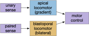
The chimera model posits that brains of bilateral animals combine features from apical (unilateral) and bilateral (“blastoporal” in their terminology because they focus on zooplankton larvae). The apical mode is associated with the front of the brain, such as the hypothalamus, and its locomotion is temporally gradient based, like the tumble-and-run of bacteria. The bilateral mode is more reflexive, turning left if touched on the right, like Braitenberg vehicles [Braitenberg 1984]. Apical systems include olfactory search and phototaxis, while bilateral touch, lateral line, auditory and bilateral vision in a second system. For zebrafish one study describes multiple paths as a “high road” through Hb (habenula, apical) and a “low road” through OT (optic tectum, bilateral) [do Camo Silva et al 2018].
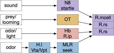
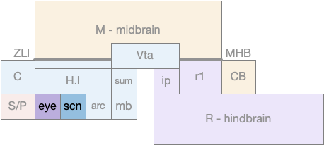
Some labeled paths for locomotion in vertebrates. H.l (lateral hypothalamus), Hb (habenula), MLR (midbrain locomotive region), N8 (acoustic-vestibular cranial nerve 8), OT (optic tectum), R.ip (interpeduncular nucleus), R.mcell (Mauthner-cell), R.rs (reticulospinal motor command)
The above diagram shows some vertebrate labeled paths, which is clearer in simpler vertebrates like the lamprey and zebrafish. In the zebrafish startle reflex, a sudden noise triggers a fast C-bend turn followed by rapid swimming. The trigger can be a noise, vestibular, or lateral line motion [Berg et al 2018]. The startle circuit is only three synapses from the original sensor to the muscle, from the N8 auditory/vestibular nerve to the giant M-cell (Mauthner cell in r4) to the motor neuron that drives locomotion. In young zebrafish larva, head touch neurons (N5 trigeminal) connect to M-cells and are later replaced by N8 [Kohashi et al 2012]. M-cells fire only once per escape to drive the initial turn. Interestingly the escape turn choice uses an axo-axonic repeater and amplifier [Guan et al 2021].
In a different path looming and dimming visual signals that represent predators or obstacles drive OT (optic tectum), which can drive escape that either uses or bypasses the M-cell depending on the threat level [Bhattacharya et al 2017]. OT also pre-programs the M-cell circuit by suppressing the left or right to avoid an obstacle [Zwaka et al 2022].
Phototaxis (seeking or avoiding light) uses a temporal gradient system composed of left Hb.m (medial habenula) and R.ip.d (dorsal interpeduncular nucleus), which projects to the R.rs (reticulospinal motor command) neurons via relays in V.mr (median raphe) and P.ldt (laterodorsal nuclei) [Chen and Engert 2014]. Food odor seeking uses the right Hb.m and R.ip.v (ventral interpeduncular nucleus) [Chen et al 2019].
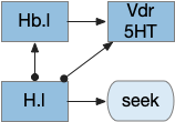
In lamprey a distinct food-seeking path through V.pt (posterior tuberculum – possibly homologous to vertebrate Vta/Snc) to MLR (midbrain locomotor region) and finally to R.rs [Derjean et al 2010]. Zebrafish has a similar dual path through Hb.l (lateral habenula) through a midbrain TSN circuit [Koide et al 2018].
Slower escape uses a distinct prepontine (rhombomere r0-r1) circuit, which is suppressed by the M-cell escape circuits [Marquart et al 2019].
Some of these paths have shared elements, particularly at the motor control like MLR, but the general pattern is multiple labeled paths for each behavior. The paths already mentioned don’t include more complex food-seeking paths through the basal ganglia and hypothalamus.
Multiple labeled paths immediately raises the difficulty of coordination. How does the system juggle priorities? Even the simple startle reflex needs to be modulated because the animal shouldn’t startle if the loud sound is expected, such as near a waterfall. In contrast in a dangerous area with possible predators the animal should increase the reflex to a hair-trigger. Similarly if the threat is weak and the animal is hunting or eating and hungry, it might ignore the threat to continue eating. A second architecture, distinct from the label path, emphasizes the coordination of multiple paths, possibly using a consensus system to decide on an appropriate action.
Consensus loops
While the labeled paths have strong evidence, the consensus loop is only a thought experiment to tie the paths together. Multiple paths for food seeking and for avoiding is a distributed system, and distributed systems makes decision circuitry more complicated because they’re not central decision node. Every node needs to agree with the decision. Whether to avoid or approach needs to be agreed on by all the systems. It wouldn’t make sense for one system to believe the animal is approaching an object but another system believes the action is avoiding. Voting distributes the consensus.
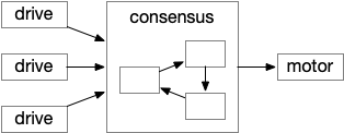
Illustration of a consensus loop. Multiple drives or labeled paths vote for approval to drive motor output.
The above diagram shows the model. The different labeled paths of seeking or avoiding join a voting consensus system in a motivational look, which allows one path to drive motor output.
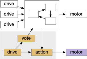
Consensus system showing one path. The driving sense or motivation tries to disinhibit itself by voting in the consensus loop.
A single labeled path has a sense or motivation drive that tries to act on motor output, but is inhibited by the consensus system. For example, if a predator odor arrives, the odor avoidance path votes to enable its own locomotion. If the consensus system agrees, it will disinhibit the odor avoidance path, letting the animal escape. Note that a high priority threat could bypass the consensus system.
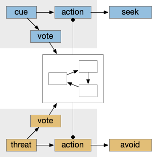
Seek and avoid consensus system
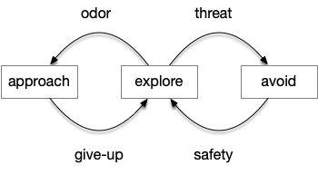
This system can manage conflicts between seek and avoidance, such as animals continuing to eat if a predator threat exists but is low. Consider a simplified consensus system with only one seek node and one avoid node, using the consensus to select one when there’s a conflict.
Managing conflicts between seeking and avoiding.
If there’s a food cue and no conflicting threats, the food vote passes easily and the animal seeks the food. Similarly a predator odor with no conflict will enable avoidance. If there’s a conflict, the system can weigh the costs and benefits of the threat and the food, possibly depending on hunger state or a more sophisticated threat assessment.
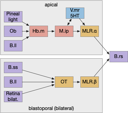
Keeping these general ideas of the labeled path and consensus systems in mind, let’s start working through several specific paths. The end goal is to organize the main brainstem locomotive areas into a simplified, unified model. The two major paths will be apical paths through Hb (habenula) using temporal gradients (klinotaxis) [Chen and Engert 2014] and bilateral paths through OT (optic tectum) using spatial gradients (tropotaxis).
Apical and bilateral avoidance
Because there are many labeled paths, dividing them up might help organize the model. An early division between labeled paths goes back to the bilaterian (worm-like, slug-like) ancestors, which added bilateral, dual-sensory navigation (tropotaxis, spatial gradient) to an older single-sensor navigation that used the animal’s movement to choose a direction (klinotaxis, temporal gradient), such as the simple tumble-and-run that even bacteria and simple radial zooplankton use for seeking odors (chemotaxis) and seeking or avoiding light (phototaxis). This chimera hypothesis [Tosches and Arendt 2013] considers bilateral animals as a fusion between the locomotive systems. The apical zooplankton larvae of bilaterian worms may have been a secondary development to escape predation [Mallatt 2021]. In vertebrates, apical klinotaxis is implemented by Hb (habenula) temporal gradient seeking and H.l (lateral hypothalamus) motivation. Bilateral tropotaxis navigation is implemented by several labeled path systems, typified by OT (optic tectum) and the M-cell start reflex.
A primitive apical example is the helical phototaxis of many annelid (marine worm) zooplankton larvae, and a primitive bilateral example is the mollusk sea slug navigation.
Zooplankton apical phototaxis
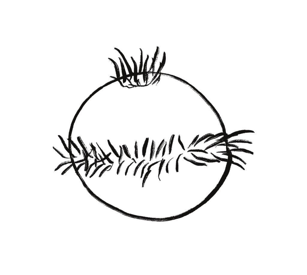
One type of zooplankton is essentially a globe with a fringe of cilia and an apical tuft for chemical processing, such as the Platynereis larva.
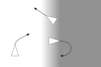
Phototaxis for this larva depends on its helical movement (helical klinotaxis). As it moves forward, the larva also rotates and wobbles, which means that parts of the equatorial band are nearer the light or further from the light depending on the rotation. If the upper cilia halt, the larva will steer toward the light. If the lower cilia halt, the larva will steer away from the light [Randel and Jékely 2016].
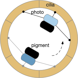
Phototaxis for an zooplankton larva.
The system depends on a directional eye, which uses a photoreceptor and a pigment cell that imposes directionality by shading the photoreceptor, because other cells of the larva are transparent. The photoreceptor compares the current brightness to its average brightness as the larva rotates. If it’s brighter than average, then it must be facing the light, and will signal the cilia to briefly halt, using ACh (acetylcholine) as a neurotransmitter. This trivial one-neuron circuit is sufficient for simple phototaxis [Randel and Jékely 2016].
Although this example larva uses two photoreceptors, it’s not truly bilateral and the two photoreceptors don’t communicate. Ablating one photoreceptor doesn’t abolish phototaxis, although it does reduce efficiency. Using three or four photoreceptor/pigment pairs would work, as well as removing all but one. This system is apical klinotaxis, not bilateral tropotaxis, which makes sense because the above zooplankton is not bilateral. While this zooplankton uses helical klinotaxis, another common form of klinotaxis is a side to side “casting” motion used by other simple animals like c. Elegans [Izquierdo and Lockery 2010].
If zooplankton phototaxis is an example of apical navigation, then the mollusk sea slug is an example of bilateral navigation.
Mollusk sea slug seek and avoid
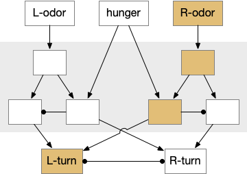
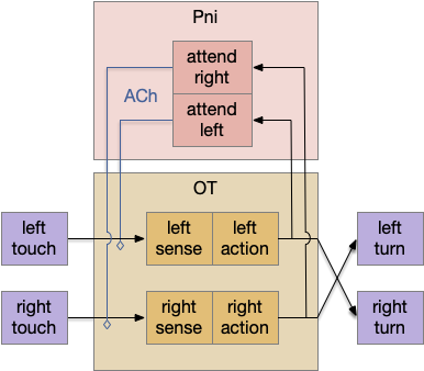
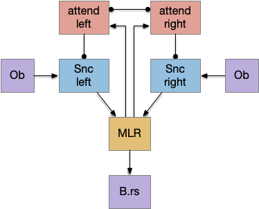
The mollusk sea slug circuit is a pure bilateral circuit, almost directly a Braitenberg circuit [Braitenberg 1984], discussed in essay 14. The following shows a rough schematic of the sea slug seek and avoid. This circuit is interesting because with only a few neurons, the slug can switch from turning toward a food odor when hungry to turning away from the odor when not hungry [Gillette and Brown 2015].
Odor seek and avoid circuit for a sea slug. Hunger switches a food odor from seek to avoid.
In the diagram above, the central grey area is a switchboard circuit. Hunger reconfigures the switches connecting the odor to the turn motor neurons. When the slug is hungry, the right odor sensor connects with the left turn muscle, seeking the odor. But when the slug is sated, the right odor sensor connects with the right turn muscle, avoiding the odor. When the slug is hungry, it approaches food but when it’s not hungry, it avoids food odor cues.
A similar animal with a different circuit configuration uses serotonin to switch from avoidance to approach [Hirayama et al 2014].
For the goal of this essay, avoiding a failed food cue, this circuit is perfect because when the animal finds a false cue, it reversed movement from seek to avoid, which exactly fits the essay needs. Unfortunately, the vertebrate circuits aren’t nearly as straightforward. As a start for the vertebrate navigation paths, the startle reflex managed in vertebrates by the giant Mauthner cells is a simple starting point.
Amphioxus fast twitch reflex
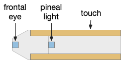
The fast twitch startle reflex is a clear example of a bilateral labeled path avoidance circuit. A noxious sense on one side causes a fast turn away from the sense. The sense can be a touch on the head, such as running into an object, or a loud sound or a vestibular imbalance signal. This circuit predates vertebrates and a similar circuit exists in amphioxus, a filter-feeding chordate that looks something like a fish without a distinct head and without eyes, but with several photoreceptors including a frontal “eye.”
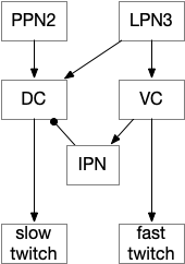
In amphioxus the startle reflex drives fast twitch muscle fibers, where normal swimming uses slow twitch fibers [Lacalli and Candiani 2017]. This circuit path is entirely distinct even to using the different muscles. The following diagram shows part of the amphioxus motor control circuit. (Because the neuron names are specific to amphioxus, they’re not hugely important for this essay.)
Amphioxus fast twitch escape uses LPN3, glutamate large paired neuron.
The diagram shows the LPN3 (large paired neuron) fast twitch escape path, and the PPN2 normal swimming match, including intermediary motor control neurons [Lacalli and Candiani 2017]. This amphioxus escape circuit resembles the zebrafish Mauthner cell escape.
Zebrafish Mauthner cell escape
Zebrafish have a pair of large M-cell (Mauthner cell) neurons that are specialized for auditory and vestibular startle escape. These are very fast reflexes on the order of 10ms, which can be modulated by higher context [Zwaka et al 2014] including OT. Although the M-cells perform a similar role to the amphioxus LPN3, it’s not clear that they’re homologous, which requires common descent, because the large escape neuron is a common pattern in non-chordate systems.
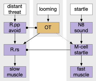
Zebrafish startle response at right in context with other labeled paths. M-cell (Mauthner R.rs cells in r4), N8 (acoustic/vestibular cranial nerve 8), OT (optic tectum), R.pp (prepontine avoidance in r0-r1), R.rs (reticulospinal motor control)
The primary input to M-cell escape is an auditory and vestibular signal from N8 (8th cranial nerve is auditory and vestibular). In water, sound and primitive vestibular sense have some similarities, because water motion produces not just sound but animal motion, depending on the frequency. The M-cell directly connects to motor neurons to muscles. The startle escape is only a three neurons and a clear, distinct labeled path.
A second zebrafish threat avoidance path uses neurons in R.pp (pre-pontine r0-r1) [Marquart et al 2019] for more distance threats. Unlike the M-cell circuit, this R.pp path is more than a reflex, but it’s still a hardwired path. A third threat circuit uses OT (optic tectum), for example the looming response. Most vertebrates will flee or freeze from a rapid and overhead expanding dark object, representing a potential predator or an obstacle. The mammalian startle circuit shares similarity with an acoustic projection to R.pn.c (caudal pontine reticular) neurons, in an analogous area to the M-cell [Kim et al 2017].
Some of these circuits do share sub circuits. For example, hindbrain locomotion and turning are distinct circuits that are used by both bilateral and apical avoidance circuits.
Hindbrain locomotion and turning
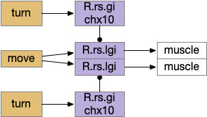
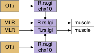
Senses are not the only source of distinct paths because actions can be split into parts like a car’s divided steering and acceleration. In vertebrates, accelerating and turning use distinct hindbrain circuits. Although both MLR (midbrain locomotive region) and OT.d (deep layer of the optic tectum) encode seeking and avoiding, they don’t encode left or right turns. Activating the left or the right MLR produces straight movement [Brocard et al 2010]. Turning is managed from OT.i (intermediate layer of the optic tectum) to distinct R.rs motor command neurons, marked by the chx10 transcription factory [Cregg et al 2020].
Hindbrain acceleration and turning circuits. R.rs (reticulospinal motor control)
The above diagram shows the basic idea. The upstream MLR can command forward movement without specifying details, because swimming is an oscillatory process with CPG (central pattern generators) in the spinal cord and the hindbrain. To turn, the chx10 neurons inhibit the swimming stroke in one direction [Cregg et al 2020], similar functionally to the apical zooplankton inhibition of cilia for phototaxis.
Splitting out turning can simplify the system by dividing labor, where OT.i is always responsible for obstacle avoidance, but a diverse set of labeled paths decode whether to seek or to avoid.
Optic tectum and dimming
The OT is named after its retinotopic visual map that is used for avoiding looming/dimming obstacles and predators, and also for seeking prey [Basso et al 2021]. For most vertebrates, OT is the primary visual area, and the visual cortex only provides abstract context, and amphibians and fish lack a proper visual cortex [Heap et al 2018]. For this essay, OT is less important for its sophisticated visual organization, but more because it also contains motor maps for seeking or prey and avoidance of looming objects, and dimming fields. Its motor map also contains drinking and licking [Liu et al 2022].
Looming/dimming path through optic tectum. OT.m (medial, deep optic tectum), R.rs (reticulospinal motor command)
OT processes looming and dimming objects and avoids them. Since the essay’s model lacks proper vision, the dimming is currently most important. Because OT also has obstacle avoidance, it’s a more sophisticated system than simply reflex. It’s likely that other avoidance systems will use OT for obstacle handling. Even in the case of the M-cell reflex, the OT.i pre-programs the M-cell, to avoid obstacles in case of a future startle [Zwaka et al 2014].
Optic tectum and turning
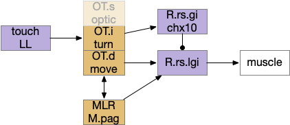
This division between turning and acceleration applies to OT itself. OT is a layered structure where the top layer is a visual map, the intermediate layer integrates other senses and produces turns, and the deepest layer includes actions such as avoiding and seeking [Liu et al 2022]. OT.d (deep OT) is a motor area for seek and avoid, connected with MLR and M.pag (periaqueductal grey) motor output, and integrates general dimming from the retina with distinct expansion calculation in OT itself [Heap et al 2018], to avoid looming objects. OT.i (intermediate OT) includes multi sensory integration and turning motor area, connected with LL (lateral line) electro sensation and water motion, somatosensory (whiskers in mice), auditory input from M.ic (inferior colliculus) and optic input from OT.s (superficial OT).
Optic tectum layered structure, emphasizing turning and motion. LL (lateral line water motion), MLR (midbrain locomotor region), OT.s (superficial optic tectum), OT.i (intermediate OT), OT.d (deep OT), R.rs (reticulospinal motor command)
Because only the top layer is specifically optic, some neuroscientists use “tectum” (roof in latin) instead of OT to emphasize its multi sensory and motor function, not just the optic features. On argument suggests that the optic layer OT.s is a secondary layer, added to a more primitive OT.i and OT.d that are more connected with reticular areas like MLR and M.pag [Edwards 1980], [Basso et al 2021]. With that argument, OT is primarily a moving and turning structure, receiving turning and moving input from touch, lateral line, primitive dimming, and other directional senses and combining with seek and avoid decisions. When the visual system developed enough detail to support crude images like looming disks or moving prey-like dots, the OT integrated vision into its top layer.
On the other hand, since OT.d receives dimming information from H.lg (central lateral geniculate nucleus) for looming escape [Heap et al 2018], it’s also conceivable that the base OT function is visual escape from dimming, where the later expanding, looming visual processing is an optimization.
Optic tectum obstacle avoidance combined with MLR seek or avoid movement. MLR (midbrain locomotor region), OT.i (intermediate optic tectum), R.rs (reticulospinal motor neurons).
This separation of obstacle avoidance turning from seeking and avoiding greatly simplifies some other circuitry that doesn’t need to duplicate the obstacle avoidance. Since other circuitry from the apical path, like the Hb-R.ip (habenula – interpeduncular nucleus) has its own turning system, OT.i doesn’t have a monopoly on turning. But even in that case, OT.i obstacle avoidance can inform apical navigation.
Some of these avoidance circuits are from the bilateral part of the chimera, such as the M-cell and the looming OT circuits, and others are from the apical part, such as Hb.m phototaxis, chemotaxis, and thermotaxis. So, let’s now more from the bilateral avoidance circuits, explore the vertebrate apical navigation.
Tunicate helical swimming and phototaxis
Tunicates (including sea squirts) are the closest chordates to the vertebrates, but because they have evolved at a greater rate and in specialized directions, comparison with vertebrates is difficult [Stolfi and Brown 2016]. Ascidian tunicates (sea squirts) have a mobile tadpole stage that plants itself in under 24 hours and transforms into a sessile filter feeder, reforming the entire brain. In general, neuroscientists believe amphioxus more resembles the ancestral vertebrate, and that ascidians have lost too many ancestral structures for a reasonable comparison [Holland 2016]. But for the sake of exploration let’s run through a thought experiment as if the ascidian larva is a compressed and simplified version of the vertebrate ancestor, although possibly only the vertebrate larva.
Specifically, consider phototaxis in the apical helical klinotaxis mode that follows a temporal gradient, since both amphioxus and ascidian larva swim in a helical pattern. Even bacteria can follow odor gradients [Hengenius et al 2012] and as discussed above zooplankton phototaxis can move toward light with only a single photosensor [Randel and Jekely 2016]. Both amphioxus and ascidian larva have single unpaired eyes, amphioxus as a single frontal eye [Lacalli 2022] and ascidians with an asymmetrical eye paired with a second pigment cell used for geotaxis as a primitive vestibular sense [Hoyer et al 2024]. In both cases, the “eye” is directional with a pigment cell, but a non-image-forming collection of photoreceptors. The ascidian asymmetrical eye works because the ascidian tadpole swims in a helical pattern so the timing of the light on the eye matters more than its position [Ryan et al 2016].
Ascidian larvae swim in a helical pattern comprised of unilateral tail flicks and symmetrical swimming [Ryan et al 2017] and use the asymmetry of the photoreceptor and photopigment to swim toward light [Mast 1921], [Zega et al 2006]. Since helical swimming doesn’t need stabilizing fins or vestibular systems to manage roll, yaw, and pitch with 3d swimming, it’s available to evolutionarily simpler systems. Another advantage of helical phototaxis is that the photoreceptors are auto-calibrating by simply averaging the light in a rotation and requires less circuitry than a bilateral comparison of light [Randel and Jekely 2016].
However, unlike the trivial zooplankton circuit that directly connected the photoreceptor to arrest the cilia, ascidian larvae need to modulate the bilateral swimming in the primitive hindbrain, timing the muscle inhibition to achieve the same effect.
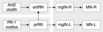
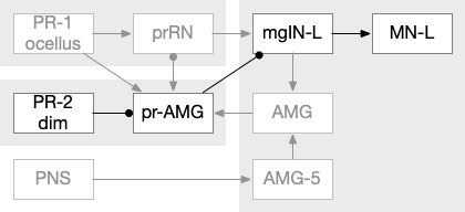
The ascidian ocellus (“eye”) has two types of photoreceptors with distinct responses. Type 1 has a pigment and lens and is directional (37 cells), while type 2 is non-directional (no pigment partner) [Salas et al 2018]. If the pigment is genetically deleted, the animal can’t use phototaxis but does respond to dimming with an escape response. In other words, the dimming response and phototaxis use distinct labeled paths with distinct input neurons [Kourakis et al 2019]. The following shows the circuit for the type 1 photoreceptors for phototaxis, where the boxes represent single neurons or small collections (5-8) of neurons, not large functions (from [Ryan et al 2016]).
Ascidian larva phototaxis (ocellus) and geotaxis (otolith) circuit. Ant2 (antenna geotaxis sensors), antRN (antenna relay neuron), mgIN (motor ganglion interneurons, left and right), MN (motor neurons, left and right), PR-1 (type-1 phototaxis photoreceptors), prRN (photoreceptor relay neuron)
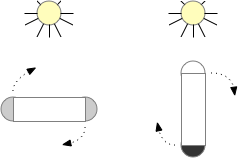
The above diagram shows both geotaxis and phototaxis circuits, which are specific to right or left motor neurons respectively, because the ascidian larva neuron circuits are highly asymmetrical. Ascidian larva geotaxis swims upward and phototaxis swims away from light, generally downward. The combination encourages swimming to the underside of ledges, such as the underside of boats and harbor piers [Ryan et al 2016]. Because of the helical swimming, the left and right motor neurons aren’t left or right turns, but turns toward or away from the target. Although this circuit is more complicated than the purely apical zooplankton because of the interface to bilateral swimming, the helical swimming keeps the circuit relatively simple.
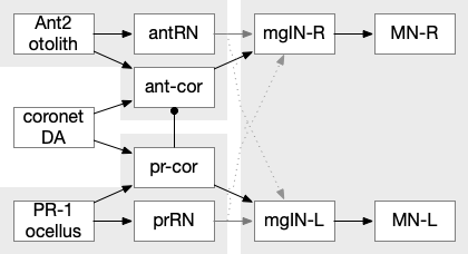
The above partial circuits are complicated by the coronet cells, another sensory cell that are paired with the photoreceptors, but with unknown function. The circuit connectivity is interesting, because coronet cells modulate both the phototaxis and geotaxis paths, but aren’t a path of their own. The phototaxis and geotaxis relay neurons above are partially bilateral. Only 70% of their connectivity is to the main side, but 30% of the connectivity is to the opposite side. In contract, the coronet-enabled neurons are 100% to the main connection [Ryan et al 2016].
Coronet cell modulation of phototaxis and geotaxis in the ascidian larva. ant2 (antenna geotaxis cell), ant-core (antenna-coronet relay neuron), antRN (antenna relay neuron), DA (dopamine), mgIN (motor ganglia interneuron, left and right), MN (motor neuron, left and right), PR-1 (photoreceptors type-1), pr-cor (photoreceptor-coronet relay neuron), prRN (photoreceptor relay neuron)
As a thought experiment (unsupported by scientific evidence) the main phototaxis path might be uncertain and stochastic, while the coronet-enabled path would be a certain, deterministic connection. If the coronet cells measured the certainty of the animal’s current direction, it could encourage sticking to the current path. For example, if the coronet cells were food-odor gradient sensors, they could fire when the animal was heading toward food, enabling a chemotaxis based on modulation of geotaxis and phototaxis.
Tunicate dimming response
The ascidian dimming response triggers locomotion with a strong turn as an escape response to predators [Kourakis et al 2019]. Unlike the phototaxis photoreceptor, the dimming photoreceptors are non-directional because’er not shaded by the pigment cell. There are 23 type-1 directional photoreceptors and 7 type-2 non-directional photoreceptors for dimming.
The above diagram shows the dimming path in context with the previous phototaxis path. Like the phototaxis path, the dimming path starts from the type-2 photoreceptors to a relay neuron and to the control neurons in the motor ganglion. Unlike the phototaxis path, the dimming path is modulated by ascending motor signals from AMG (ascending motor ganglion) and from the phototaxis path [Ryan et al 2016], presumably so the normal helical phototaxis doesn’t trigger a dimming response.
Cement gland and attachment
The ascidian larva hatches before dawn, swims upward for a few hours because geotaxis is enabled before phototaxis neurons attach, and then swims away from the light, settling on a lively rock, preferring a ledge to settle under if possible. Larva do not feed [Ryan et al 2016]. The larva will attach with a cement gland on the front of its head, a trio of palms, and then transforms into the adult sessile filter feeder. The palp sensors trigger the attachment circuit, which stops all swimming and begins the metamorphosis [Anselmi et al 2024].
Although the full details of the above circuit [Ryan et al 2016] aren’t critical, the PN (palp neuron) senses the animal bumping into a rock modulated by chemical senses that avoid toxic area, and triggers a swimming shutdown by inhibiting the motor neurons and interneurons [Hoyer et al 2024]. Like the previous diagrams, the boxes represent individual neurons or small group, not large functional regions.
While the ascidian cement gland is permanent, several fish [Pottin et al 2010] and amphibians [Rétaux and Pottin 2011] have a homologous cement gland used for larvae, not adults. For example, frog tadpoles can attach to the bottom of leaves or to the water surface to avoid predators until they are large enough to hunt [Jamieson et al 2000], [Yoshizawa et al 2008]. Because of the widespread cement gland among many fish species and amphibians as well as the tunicates, it’s likely the original vertebrates had a similar cement gland [Rétaux and Pottin 2011]. Whether the gland was larva-only like in vertebrates or also used for adults as in tunicates is unknown. In either case, the cement gland circuit that inhibits locomotion must have been part of the original vertebrate.
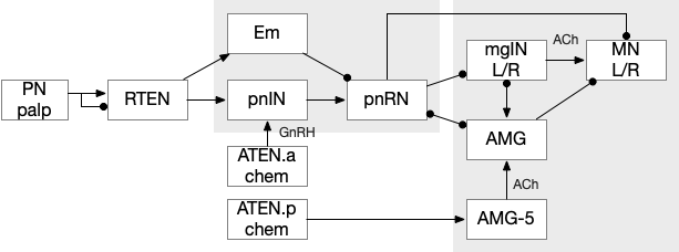
Vertebrate analogies to the ascidian circuits
Because the ascidians are so specialized and reduced from the common ancestor with vertebrates, including major losses in genes, cells and structures, comparing the two is essentially impossible to be homologous (shared descent) [Holland 2016]. However, for the sake of exploration, I’m ignoring that advice, and looking for analogous vertebrate circuits to the ascidian larva.
The ascidian behavior each have distinct circuit paths that mostly only come together at the motor control neurons. The exception is the feedback from the AMG (ascending motor ganglion) neurons, which do feedback to the midbrain neurons, but the main paths are separate forward paths. Each of the geotaxis, phototaxis, dimming, cement gland attachment, and bilateral escape are circuit paths that are distinct until the motor command neurons.
A vertebrate analogy to the ascidian phototaxis gradient path might be the path from the retina to Hb.m (medial habenula) to R.ip (interpeduncular nucleus) and V.mr (median raphe), which then project to R.rs (reticulospinal motor command). Like the ascidian path, the Hb-R.ip phototaxis path is relatively isolated from the other paths, although Hb.m does receive large modulation from the hypothalamus. Although R.ip is mostly descending, like ascidian mgIN, V.mr is both ascending and descending like mgIN and AMG.
The dimming path from the type-2 photoreceptors resembles the dimming input to the vertebrate OT (optic tectum). Although existing vertebrates have more sophisticated eyes that can distinguish expanding objects, the dimming input to OT is still important and used for escape directionality [Fotowat and Engert 2023], [Heap et al 2018]. Retina dimming cells reach AF6 and AF8 [Temizer et al 2015], which are thalamic arborization fields before reaching OT.d. Although more complicated expanding looming response in vertebrates is better studied, expansion detection requires an image-supporting eye, and OT.d receives the simpler dimming input. Like the ascidian dimming pr-AMG (photoreceptor – ascending motor ganglion) neuron, OT.d receives multiple ascending and descending inputs that modulate the dimming response. In particular Ppt (pedunculopontine nucleus) and P.ldt (laterodorsal nucleus) both receive OT.d output and forward to R.rs, functionally similar to mgIN (motor ganglion interneuron), and send ascending feedback from R.rs to OT.d, resembling the AMG (ascending motor ganglion) functionality.
Because the cement gland exists in vertebrates, the circuit should be available, and studies do show that N5 (head touch trigeminal nerve) innervates it automatically [Pottin et al 2010], but I haven’t read any study that says this this specific group of trigeminal neurons connects to. As a through experiment, consider H.stn (subthalamic nucleus) as a choice for the cement gland, because H.stn halts ongoing action, and because H.stn receives direct input from C.i (insular cortex) and C.ss (somatosensory cortex), which are more sophisticated versions of the chemo / mechanosensory palp neurons.
The coronet path enhances taxis confidence, reducing stochastic choice, and is a set of dopamine neurons. The striatum circuit and dopamine’s role has a similar function. Without dopamine, the basal ganglia suppress weak input, and allow stochastic action. With dopamine, the basal ganglia suppresses the randomness and keep action on track. This path resembles rheotaxis food seeking, where a fish approaches a food odor by swimming upstream [Coombs et al 2020]. The “what” signal (odor) differs from the “how” signal (water current cues). Like rheotaxis, the coronet cells enhance the existing phototaxis and geotaxis, reducing the default stochastic noise.
Hb.m Medial habenula
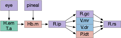
Of the ascidian labeled paths above, the Hb (habenula) phototaxis path will be a useful anchor for the upcoming consensus circuit. Like the ascidian asymmetrical phototaxis neurons, the vertebrates Hb.m (medial habenula) is also governed by Nodal asymmetry [Roussigne et al 2009], where Nodal is a developmental genetic transcription factor. In zebrafish the left Hb.m support phototaxis, and the right Hb.m supports chemotaxis [Chen et al 2019]. Hb.m phototaxis receives both “on” and “off” neurons from the retina with a relay either in H.em (pre thalamic eminence) [Zhang et al 2017] or T.a (an area in the anterior thalamus) [Cheng et al 2017], where the connections are debated. Although Hb.m does receive dimming input from the adjacent photoreceptive pineal gland, the retina photoreceptors are more important for phototaxis [Dreosti et al 2014].
As an anatomical note, the zebrafish Hb.m is actually dorsal and therefore named Hb.d. Similarly the zebrafish Hb.m is ventral and named Hb.v, but as a simplification I’ve used the mammalian name.
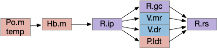
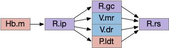
The output path from Hb.m is through R.ip (interpeduncular nucleus), which projects to several areas including R.gc (pontine central era), V.mr (median raphe – serotonin), V.dr (dorsal raphe – serotonin), and P.ldt (laterodorsal nucleus – ACh) [Quina et al 2017]. The V.mr glutamate and GABA neurons may be more important for this circuit than the serotonin neurons, which they outnumber. Also, note that V.mr is located in the same hindbrain rhombomeres (r2-r5) as some of R.rs, but are more ventral, and are reciprocally connected. In other words, V.mr is highly action and motor associated.
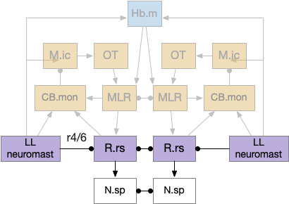
As described above, the Hb.m-R.ip path is a klinotaxis path for phototaxis [Chen and Engert 2014], chemotaxis and thermotaxis, where the klinotaxis is temporal from the animals movement, but not the helical movement of the ascidian larvae. The Hb.m-R.ip klinotaxis has multiple inputs for lamprey, including light, odor, and lateral line (water movement) [Stephenson-Jones et al 2011].
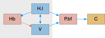
Habenula klinotaxis for lamprey for light, odor, and lateral line. Hb.m (medial habenula), LL (lateral line), R.ip (interpeduncular nucleus)
Although I’ve focused on Hb.m as an avoidance gradient circuit, it’s also a food odor seeking circuit [Chen et al 2019]. The Hb.m klinotaxis for light and odor also applies to temperature, using input from Po.m (medial preoptic nucleus) [Palieri et al 2024] and social seek and avoidance [Okamoto et al 2021], [Chou et al 2016].
Because Hb.m has several sub-nuclei and genetic clusters, it likely represents different labeled paths, supporting multiple distinct seek and avoidance paths. A binary seek vs avoid circuit is likely an oversimplification, because studies have found at least 5-6 olfactory Hb.m clusters in the larval zebrafish [Jetti et al 2014], [Beretta et al 2014]. Hb.m is asymmetrical, like the ascidian larva. Odors from either olfactory bulb activate the right Hb.m [Chen et al 2019]. Hb.m neurons have at least 262 neuropeptide receptors [Ables et al 2023] as well as morphine receptors [Gardon et al 2014], [Boulos et al 2020] including neuropeptides modulating hunger or social motivation from hypothalamic areas like H.l and H.pv.
R.ip interpeduncular nucleus
Since I’ve already covered some of the R.ip klinotaxis function in essay 24 and essay 25, I’m going to focus on the R.ip connectivity, particularly the ascending connectivity. R.ip descending efferents don’t target R.rs directly, but instead use intermediaries like R.gc (pontine central gray), V.mr (median raphe) and P.ldt (laterodorsal tegmental area) [Lima et al 2017], [Quina et al 2017].
The ascending afferents of R.ip also work through intermediaries, particularly P.ldt and V.mr [Quina et al 2017], although other connectivity studies report R.ip as directly producing ascending connectivity [Lima et al 2017]. Because V.mr is directly caudal to R.ip, the disagreement is essentially about the boundaries between R.ip and V.mr.
The ascending R.ip connectivity will become important in the next section on the consensus circuit because it completes the consensus loop, where other labeled path connectivity is descending. The ascending role is analogous to the ascidian AMG (ascending motor ganglion) neurons. For a consensus circuit to work, all nodes need to be informed of the consensus decision.
Consensus circuit narrative
Let’s now consider the consensus circuit and how it might develop from a strict labeled path system. This is just a thought experiment as a narrative explanation for the Hb.l (lateral habenula) system.
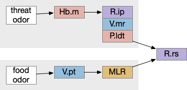
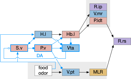
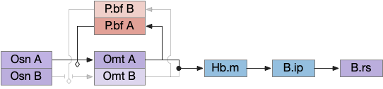
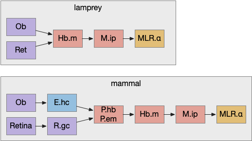
For simplicity, let’s restrict the narrative to apical systems only, ignoring bilateral systems like OT, and let’s start from a labeled path system. In the lamprey, odor information from Ob (olfactory bulb) splits into multiple paths. One path reaches Hb.m directly and another contacts V.pt (posterior tuberculum), considered a homologue of Vta / Snc (substantia nigra pars compacts), which then contacts MLR in lamprey [Derjean et al 2010] and zebrafish [Kermen et al 2013].
In this example, these two paths are distinct with threat odors going through the Hb.m – R.ip circuit and using P.ldt as an apical version of the MLR (which in lamprey may not be distinct from Ppt MLR, since the lamprey doesn’t have distinct Ppt, P.ldt and M.cnf (cuneiform nucleus)). The animal seeks food using the V.pt to MLR path.
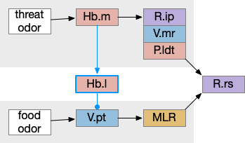
These two system can come into conflict. For the above simple system, suppose conflicts are resolved in R.rs itself, as a hard-coded priority where threats always win. But now consider a system where the conflict is resolved earlier in the stream by adding Hb.l as a lateral inhibition relay.
As a first step consider lateral inhibition of threat odor suppressing food seeking. Here the lateral inhibition path uses a relay from Hb.m to Hb.l [Gouveia and Ibrahim 2022] in a primitive Hb.l that then suppresses the V.pt path. This lateral inhibition duplicates the earlier lateral inhibition in R.rs, but is more specific because it inhibits earlier in the two paths.
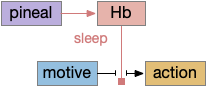
In the above diagram, the blue lines represent new connections. Notice the gating pattern for V.pt resembles the gating for the consensus circuit where the action nodes are V.pt and V.mr. Hb.l then becomes the vote accumulator for the consensus circuit. Also notice the similarity with the sleep model from essay 29, where Hb inhibits food seeking for sleep. An alternative narrative might repurpose the sleep inhibition into a path inhibition [Hikosaka 2010].
Both Hb.l and Hb.m are tonically acting, meaning that without any input their resting output is a middle value, not a binary output. This means Hb.l can gate V.pt seek and also gate its opposing avoidance circuit in V.mr and P.ldt.
For the next step, let’s add both a bidirectional selection and also add some internal state management, because the animal shouldn’t seek food if it’s sated.
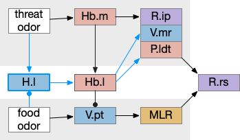
H.l hunger modulation
H.l (lateral hypothalamus) has access to hunger and satiety information by sensing blood levels directly and from connections from R.pb (parabrachial), which has signals from the digestive system via N10 vagus nerve through R.nts (solitary tract nucleus). When Ob (olfactory bulb) senses a food odor , H.l can modulate it with the current hunger sense. This means H.l as gating input to Hb.l is more effective than the simple lateral inhibition from Hb.m If the animal is sufficiently hungry, it might ignore weak threats. Note the similarity to the mollusk sea hare circuit, where hunger changed food odor from seeking to avoidance depending on the internal state.
Adding H.l hunger modulation to the decision between the threat odor avoidance path and the food odor seek path. H.l (lateral hypothalamus), Hb.l (lateral habenula), Hb.m (medial habenula), MLR (midbrain locomotor region), P.ldt (laterodorsal tegmentum), R.ip (interpeduncular nucleus), R.rs (reticulospinal), V.mr (median raphe), V.pt (posterior tuberculum).
In addition to hunger, other internal states can modulate Hb such as hypothalamic threat signaling ([Wagle et al 2022]. This step also adds control of the threat path, taking advantage of the Hb.l tonic activity to either inhibit food seeking or threat avoidance.
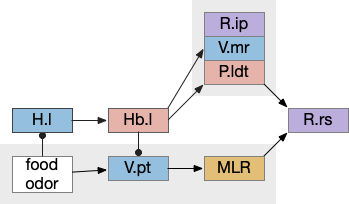
Place avoidance without a threat
Suppose we take the above circuit, but ignore or disable the threat avoidance path via Hb.m. Even without the threat path, there is an avoidance path from Hb.l to V.mr and P.ldt, where Hb.l not only disinhibits threat avoidance, but can produce place avoidance without a threat.
The above diagram shows deletion of the threat path, while retaining the abstract place avoidance path. If place avoidance is triggered, the animal will avoid the current location without needing a specific threat to avoid. This means that H.l stimulation by itself can trigger real-time avoidance [Stamatakis et al 2016]. In mammals the H.l to Hb.l connection has at least 6 clusters [Calvigioni et al 2023], which suggests multiple paths even in the abstract place avoidance.
S.v ventral striatum digression
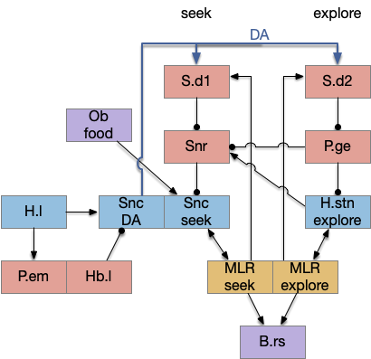
This model of the seek vs avoid circuit can be extended to S.v (ventral striatum aka nucleus accumbens) and P.v (ventral pallidum). Consider S.v / P.v as a generalization of H.l, providing more general context beyond hunger. This basal ganglia extension allows for a positive feedback loop. which enables multiple rounds of voting, integrating values, such as with drift diffusion.
In the above, I’ve split the V.pt of the lamper into an ascending Vta (ventral tegmental area) dopamine area from mammals, but left the V.pt to represent the descending glutamate / GABA portion of Vta, despite mammals lacking a distinct V.pt. If there’s a food cue when hungry, H.l to Vta stimulation will generate high DA in S.v, enabling it, which will disinhibit V.pt to enable food seeing. Here, S.v / P.v is acting as the consensus circuit and the V.pt path is the action for food seeking.
As with the smaller Hb.l circuit, S.v / P.v is also part of a sleep / wake circuit using dopamine as a wake signal, as used in essay 29. If the animal is currently seeking food, it shouldn’t fall asleep, and the high dopamine signals to stay away. Again, from a narrative sense, this circuit could have been repurposed from a wake circuit, as opposed to a path conflict system.
In zebrafish Hb.l only projects to V.mr and does not project to any DA [Amo et al 2014], while in the more primitive lamprey Hb.l projects to both V.mr and DA [Stephensen-Jones et al 2011], which suggests that the V.mr projection is more functionally critical to this circuit than the Vta projection, or that the Vta circuit is a later development. The zebrafish V.pt has descending dopamine but the existence of significant projections to the striatum is questioned [Yamamoto and Vernier 2011].
Note that H.l retains its central role, where the S.v circuit generalizes the base H.l function without replacing it. Stimulating H.l.g (H.l GABA neurons) can trigger seeking through its projection to Vta [Nieh et al 2016], and stimulating H.l.glu (glutamate H.l neurons) can trigger place avoidance through the H.l.glu projection to Hb.l [Stamatakis et al 2016].
Hippocampus digression
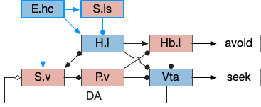
For place preference and place avoidance E.hc (hippocampus) plays a natural because E.hc represents context and place such as place cells, and H.hc projection strongly to both the hypothalamus and S.v. If we add the H.hc projections to H.l via S.ls, the seek / avoidance circuit looks something like the following.
Hippocampus modulation of H.l place seek and avoid. DA (dopamine), E.hc (hippocampus), H.l (lateral hypothalamus), Hb.l (lateral habenula), P.v (ventral pallidum), S.ls (lateral septum), S.v (ventral striatum), Vta (ventral tegmental area).
H.l has neurons that represent food zones and non-food zones [Jennings et al 2015], presumably using E.hc place information, although possibly using P.bst (bed nucleus of the stria terminals) as an intermediary.
H.sum completing consensus loop
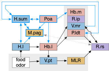
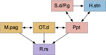
The consensus circuits needs to return the final action and motor choice back into the early layers, otherwise the motivation circuit wouldn’t know if a lower-level startle or OT looming escape took priority of the seek path. With analogy to the ascidian larva, this role resembles the AMG (ascending motor ganglia) neurons, which I associated with P.ldt and V.mr. For this consensus narrative, I’m taking H.sum (supramammillary) as the primary feedback node with an assist from Poa (preoptic area) to complete the loop to Hb.m and M.pag (periaqueductal gray).
H.sum as completing the consensus loop, linking the habenula output back to habenula input. H.l (lateral habenula), H.sum (supramammillary nucleus), Hb.m (medial habenula), MLR (midbrain locomotor region), M.pag (periaqueductal gray), P.ldt (laterodorsal tegmentum), Poa (preoptic area), R.ip (interpeduncular nucleus), R.rs (reticulospinal), V.mr (median raphe), V.pt (posterior tuberculum).
H.sum has several sub circuits with different functions, which studies are only starting to untangle. H.sum tac1 (neurotransmitter aka substance P) is strongly associated with upcoming locomotion [Farrell et al 2021]. H.sum’s Poa projection is specifically associated with threat avoidant locomotion [Escobedo et al 2023].
V.mr and P.ldt are connected with R.rs and the bilateral OT circuit, and therefore have information about the selected action at the level of the hindbrain and motor afferent copies. Both are strongly connected to H.sum. H.sum also connects with M.pag (periaqueductal gray) and H.sum activates when M.pag.d is stimulated [Pan et al 2004]. H.sum also activates when the H.vm (ventromedial hypothalamus) threat nuclei are stimulated.
H.sum is immediately rostral to Vta and highly connected with it (not shown in the diagram.) H.sum contains some DA neurons itself, which are sometimes considered as an extension of A10, the Vta dopamine neuron area, although the neuron types differ [Yetnikoff et al 2014], [Menegas et al 2015].
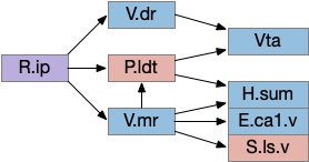
H.sum is strongly connected with E.hc (hippocampus) and is one of the few external input to both E.dg (dentate gyrus) and E.ca2 (cornu ammonia), and is a major theta source to P.ms (medial septum), which drives E.hc theta. Its link to E.hc are important for both novel object exploration [Chen et al 2020], [Takahashi et al 2023] and social memory [Qin et al 2022]. Although I’m not yet adding E.hc to the essays, the novel object detection will be important soon to avoid repeated exploration of the same object.
Note that Poa has already participated in the Hb.m to R.ip circuit because Poa drives thermotaxis [Palieri et al 2024] as part of the original Hb aversive apical path.
M.pag tetrapod complications
In a sense, the vertebrate brain is designed around fish navigation, exemplified by the simple M-cell startle circuit that requires only three neurons between the acoustic sense and the swimming muscles. Although the direct Braitenberg-like connections to R.rs work for fish locomotion, tetrapod locomotion is more complex. M.pag (periaqueductal grey) is a central grey area surrounding the midbrain ventricle (“periaqueductal”), and it an inner ring to OT, which is immediately dorsal to it. Naming it “OT.dd” (deep, deep layer of OT) would not be unreasonable. Among other tasks like vocalization [Jürgens 1994] and hunting [Marín-Blasco et al 2020], M.pag provides a similar to R.rs but at a higher level, like syllables to phonemes. So in the following examples, M.pag can be viewed as similar functionality to R.rs.
Unlike R.rs, M.pag can access more sophisticated navigation. Where the M-cell can only turn left or right, M.pag can use OT for obstacle avoidance and even higher navigation of the hippocampus using H.pm.d (dorsal premammillary nucleus) [Wang et al 2021].
M.pag flight
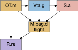
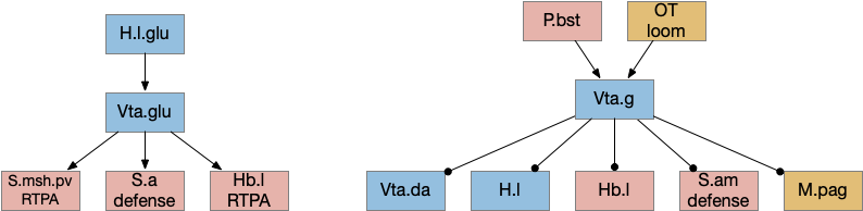
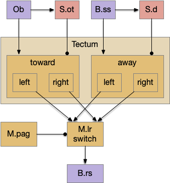
M.pag implements innate behaviors, including flight, freezing, hunting, grooming, and vocalizations. The following diagram shows some of the looming flight circuitry [Zhou et al 2019]. As before, OT.m primarily processes the looming signal and OT.m sends input to M.pag.d as an integrated threat signal, where M.pag.d computes a threshold for responding to the threat [Evans et al 2018].
M.pag flight for the looming circuit. M.pag.d (dorsal periaqueductal gray), OT.m (medial, deep optic tectum), R.rs (reticulospinal), S.a (central amygdala), Vta.g (gaba neurons of the ventral tegmental area).
In the diagram, the second interesting path is through Vta.g (Vta GABA neurons) and S.a (central amygdala). Because OT.m and M.pag.d directly output to R.rs neurons, the projects to Vta.g and S.a aren’t required for motor control, but because of the distributed consensus system, other systems need to be informed of the looming response. S.a modulates defense, hunting, and eating systems, and Vta.g also inhibits the current action by suppressing dopamine, back to the consensus loop, suppressing any current seek action.
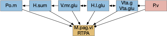
M.pag.vl avoidance
While M.pag.d is strongly associated with fast escape, M.pag.vl is more complicated with diverse functions including hunting [Franklin 2019], [Marín-Blasco et al 2020], vocalization [González-García et al 2024], and laughter [Klingbeil et al 2021]. Since this essay focuses on avoidance, where avoidance here isn’t the high speed predator escape of M.pag.d.
As discussed above, H.l is a central motivational node, filling a similar role to the central hunger node in the mollusk sea hare navigation. However, H.l is much more complicated than a simple hunger node. One developmental paper divided H.l into nine distinct regions [Diaz et al 2013], but that anatomical division understates the complexity. A genetic transcription analysis finds 15 glutamate and 15 GABA clusters [Mickelson et al 2019]. Interestingly, the Diaz study identifies their H.l.1 area with H.sum.l, treating H.sum.l as part of H.l.
In general, H.l.glu produces place avoidance and H.l.g enables seeking, but as mentioned above with at least 15 genetic types and 9 regions, this division is almost certainly an oversimplification.
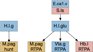
H.l seek and avoid efferents. E.ca1.v (ventral hippocampus), H.l (lateral hypothalamus glutamate and GABA), Hb.l (lateral habenula), M.pag (periaqueductal gray), S.ls (lateral septum), Vta.g (ventral tegmental area GABA).
The H.l.glu to M.pag connection is certainly capable of driving motor avoidance. Interestingly, a different H.l population is part of the M.pag hunting circuit. Both Vta.g and Hb.l enter the motivation loop. I’ve added the E.ca1.v (ventral hippocampus CA1) input to H.l because E.hc.v (ventral hippocampus) is strongly associated with place, and E.hc.v specifically with aversive context.
R.pb peribrachial nucleus
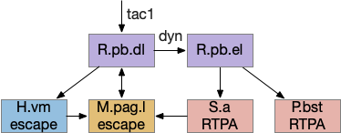
R.pb (peribrachial nucleus) is a pain, alarm, feeding, and respiration hub in the prepontine isthmus area (r0-r1). As an alarm center [Campos et al 2018], R.pb is connected with escaping and avoiding circuits. As covered in essay 29 speed, it includes a high Co2 trigger that drives place avoidance. It also includes pain triggers for escape. R.pb has multiple functions defined more by chemical markers than topology. One study explored R.pb’s role in escape and avoidance behavior [Chiang et al 2020].
R.pb avoidance circuits. dyn (dynorphin neurotransmitter), H.vm (ventromedial hypothalamus), M.pag.l (periaqueductal gray), P.bst (bed nucleus of the stria terminalis), R.pb (peribrachial nucleus), RTPA (real-time place avoidance), S.a (central amygdala), tac1 (tachykinin 1 / substance P neurotransmitter)
R.pb.dl (dorsolateral R.pb) and R.pb.el are adjacent R.pb areas that are associated with alarm and pain responses. R.pb.dl receives direct N5 (trigeminal – head, jaw) and N.sp (spinal) pain input, including pain input marked by tac1 (tachykinin 1 peptide aka substance P). Relevant to this essay, the outputs divide between direct escape behavior with not learning and indirect avoidance behavior with learning. The M.pag.l projection produces flight and jumping. The S.a (central amygdala) and P.bst (bed nucleus of the stria terminalis – extended amygdala) projections produce real-time place avoidance and are capable of CPA (conditioned place avoidance) [Chiang et al 2020]. The R.pb example is useful because it combines a direct locomotion to M.pag with output to the slower consensus circuit.
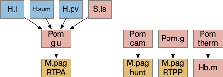
Preoptic area
Poa (preoptic area) is a multifunctional area directly anterior to the hypothalamus and often considered part of the hypothalamus, although genetic markers suggest it’s more closely related to the forebrain. Like other brainstem areas, its functionality is more organized by genetic markers than topology.
The above diagram shows some of the Pom (medial preoptic area)functions. Temperature management has been discussed with a connection through Hb.m gradient following. Threat avoidance from signals from H.sum, H.pv (periventricular hypothalamus), or S.ls (lateral septum) can lead to RTPA through a M.pag projection [Escobedo et al 2023]. Local exploration, a RTPP function, also uses a M.pag projection [Shin et al 2023], and Pom can also enable hunting [Park et al 2018], although through a M.pag projection. The recent genetic research tools will likely unravel more of its functionality.
Poa has a strong projection to both Hb.m and Hb.l, suggesting that it’s an important node in the locomotion consensus circuit. In the thought experiment I’ve outlined above, Poa is part of the feedback system through H.sum, but Poa also receives E.hc.v (ventral hippocampus) input through S.ls (lateral septum), so it may be an important node in its own right.
Ppt / P.ldt
The ACh (acetylcholine) neurons near the midbrain-hindbrain boundary Ppt (pedunculopontine tegmentum) and P.ldt (laterodorsal tegmentum) are the core of the MLR. In simpler vertebrates like the lamprey, the MLR is only a single area, generally named P.ldt. In mammals, not only are P.ldt and Ppt split, but a chunk of locomotive action is in a different nucleus M.cnf (cuneiform). Although M.cnf is more of a direct locomotive area, the locomotive neurons don’t respect the anatomical boundary, but are a group of glutamate neurons spanning from Ppt to M.ncf, where Ppt and M.cnf are neighbors [Caggiano et al 2018]. Tetrapod locomotion is more complex than fish swimming, which may be a partial reason for the expansion and division.
Ppt is strongly reciprocally connected with the deeper layers of OT: OT.i for turning and sensory integration, and OT.d for seek and avoid. Its connections resemble the R.pgb (parabigeminal aka nucleus isthmi) which sustains attention for the OT.s (superficial OT) [Knudsen 2011] and covered in essay 19. R.pgb, Ppt, and P.ldt are sibling nuclei that develop from the same area in r1 that also produces R.pb and cerebellum granule cells [Pose-Méndez et al 2023].
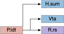
Some P.ldt connections, emphasizing that Vta connections are collaterals of R.rs. H.sum (supramammillary), P.ldt (laterodorsal tegmentum), R.rs (reticulospinal), Vta (ventral tegmental area)
P.ldt is complicated by the relative lack of recent studies of its descending projections since [Cornwall 1990] and an over-focus on its Vta connection. Because neuron tracing in [Zhao et al 2023] suggests that P.ldt has more descending connections to R.rs than Ppt and that all Vta connections are collaterals of R.rs connections, studies like [Coimbra et al 2021] and [Liu et al 2022] that find locomotion through Vta projections could be produced by its R.rs projection. P.ldt has reciprocal connections with H.sum.
Vta
Although Vta (ventral tegmental area) is most studied for its ascending dopamine projections to S.v (ventral stratum) and F.pfc (prefrontal cortex), it also contains glutamate and GABA projections, including descending connections. Non-tetrapods like fish and lamprey have a homologous V.pt (posterior tuberculum) with prominent descending locomotor connections to MLR [Ryczko et al 2017], [Derjean et al 2010]. The earlier thought experiment for the development of a locomotor consensus split out an ancient V.pt from the mammalian Vta as a way of describing the old descending functionality.
Vta glutamate and GABA connections. H.l.glu (lateral hypothalamus glutamate), Hb.l (lateral habenula), M.pag (periaqueductal gray), OT (optic tectum), P.bst (bed nucleus of the stria terminalis), S.a (central amygdala), S.am (medial central amygdala), S.msh.pv (medial shell of the ventral striatum, parvalbumin neurons), Vta.da (ventral tegmental area, dopamine), Vta.g (Vta GABA), Vta.glu (Vta glutamate)
The above diagram shows some of the connections of the glutamate and GABA Vta [Taylor et al 2014], including projections to M.pag and to Hb.l that are direct locomotor for seek and avoid. The Vta is a main dopamine source for S.v and F.pfc with multiple distinct areas. Vta.m, which projects to S.msh (medial shell of S.v) is aversive, while Vta.l, which projects to S.lsh (lateral shell of S.v) and S.core (core of S.v) promotes seek [Szőnyi et al 2019]. Vta.m is non-reinforcing, as opposed to Vta.l, which is well-studied for reinforcement.
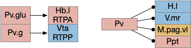
P.v ventral pallidum
P.v is a main output of S.v and the only output of S.ot (olfactory tubercle). As essay 29 covered, it’s an important sleep/wake node. For this essay, the important bit is a split between RTPP and RTPA depending on its output.
Calvigioni D, Fuzik J, Le Merre P, Slashcheva M, Jung F, Ortiz C, Lentini A, Csillag V, Graziano M, Nikolakopoulou I, Weglage M, Lazaridis I, Kim H, Lenzi I, Park H, Reinius B, Carlén M, Meletis K. Esr1+ hypothalamic-habenula neurons shape aversive states. Nat Neurosci. 2023 Jul;26(7):1245-1255.
Chen S, He L, Huang AJY, Boehringer R, Robert V, Wintzer ME, Polygalov D, Weitemier AZ, Tao Y, Gu M, Middleton SJ, Namiki K, Hama H, Therreau L, Chevaleyre V, Hioki H, Miyawaki A, Piskorowski RA, McHugh TJ. A hypothalamic novelty signal modulates hippocampal memory. Nature. 2020 Oct;586(7828):270-274.
Farrell JS, Lovett-Barron M, Klein PM, Sparks FT, Gschwind T, Ortiz AL, Ahanonu B, Bradbury S, Terada S, Oijala M, Hwaun E, Dudok B, Szabo G, Schnitzer MJ, Deisseroth K, Losonczy A, Soltesz I. Supramammillary regulation of locomotion and hippocampal activity. Science. 2021 Dec 17;374(6574):1492-1496.
Szőnyi A, Zichó K, Barth AM, Gönczi RT, Schlingloff D, Török B, Sipos E, Major A, Bardóczi Z, Sos KE, Gulyás AI, Varga V, Zelena D, Freund TF, Nyiri G. Median raphe controls acquisition of negative experience in the mouse. Science. 2019 Nov 29;366(6469):eaay8746.
The first two parts of this essay were a general overview of the necessity of sleep and some of the properties. Here I’m going over some of the brainstem circuits that control sleep.
Wake ignition
Waking requires intrinsic motivation because sleeping places the animal away from distraction, to an extreme in hibernation. A short nap, as is more typical in the waking period, needs to end without needing external stimulus or an internal one like hunger. What’s needed is an internal ignition source to drive wake and motivation.
In rodents, if the area around R.pb (parabrachial nucleus in r1) is lesioned, the animal remains in a coma [Fuller et al 2011]. For humans, a study of coma showed a pattern of the same area as consistently being destroyed [Grady et al 2022]. However, the exact cells aren’t known, and other studies that lesion R.pb for conditioned taste studies don’t produce coma. Still, this site seems a likely ignition area.
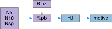
Possible wake ignition subcircuit. R.pb is the main ignition source and wakes motivational areas like H.l. H.l (lateral hypothalamus), N5 (trigeminal nerve), N10 (vagus nerve), Nsp (spinal cord), R.pb (parabrachial nucleus), R.pz (parafacial zone).
The above diagram shows a possible wake ignition circuit. The area around R.pb is the main wake ignition node. R.pb produces wake by stimulating motivational areas like H.l (and others).
It’s not known if the R.pb area is self-igniting or if astrocytes in the area are critical, or if it uses peripheral wake signals such as N5 (trigeminal nerve), N10 (vagus nerve), or N.sp (spinal cord or other periphery) [Grady et al 2022]. In the fruit fly Drosophila specialized peripheral leg neurons can promote daytime sleep [Jones et al 2023]. These specialized neurons are distinct from sensor or motor neurons. In addition peripheral neurons from Drosophila PPM area are wake promoting [Satterfield et al 2022]. So, it seems plausible that peripheral nerves such as N5, N10, or N.sp could have similar wake-producing neurons, although this is entirely speculative.
If R.pb is a wake-ignition system, then sleep needs to suppress it, whether by internal clock regulation, or external suppression. In the hindbrain, R.pz (parafacial zone near N7 and r5 / r6) suppresses R.pb to create sleep [Anaclet et al 2014]. Disabling R.pz decreases NREM sleep by 30% [Erikson et al 2019].
Hindbrain (rhombomere) sleep
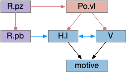
R.rs (reticulospinal) neurons in the caudal hindbrain (medulla, r6-r8) have both wake and sleep effects. Because R.rs are motor control neurons, they need to suppress sleep while they’re active, but the same area also contains sleep promoting areas. So, when experimenters stimulate the area, the animal remains awake, but immediately following the end of stimulation the animal sleeps, because the sleep-inhibition is removed. [Teng et al 2022]. These R.rs neurons (ventrolateral medulla) send collaterals to Po.vl (ventrolateral preoptic area), which is a forebrain sleep / wake area.
Midbrain sleep
A specific nucleus near N3 (oculomotor nerve) and associated motor areas (Edinger-Westphal) is a sleep promoting area. Stimulating it increases NREM [Zhang et al 2019]. (I’m just noting this for reference. I don’t understand how this area connects with other sleep areas.)
Ventral preoptic area
Although wake-maintaining areas are widely distributed, and much of sleep-circuitry is postponing sleep for ongoing actions, sleep-promoting area are more rare. One sleep-promoting area is Po.vl (ventrolateral preoptic area) and Po.mn (median preoptic area), which are adjacent area. Po.vl is inhibitory GABA and inhibits neurotransmitter wake areas like V.lc (locus coeruleous – norepinephrine), Vta (dopamine), V.dr (serotonin), M.pag.v (which is V.dr but refers to a dopamine area), and Ppt (pedunculopontine nucleus – acetylcholine) and P.ldt (laterodorsal tegmental nucleus – also acetylcholine).
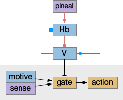
Po.vl sleep-promoting area suppresses wake-promoting areas. H.l (lateral hypothalamus), Po.vl (ventrolateral preoptic area), R.pb (parabrachial nucleus), R.pz (parafacial zone), V (wake-neurotransmitter areas including dopamine, histamine, serotonin, acetylcholine, orexin).
In the diagram above, Po.vl promotes sleep by inhibiting wake-promoting areas, here represented by H.l and V, where V includes the neurotransmitter wake areas. This Po.vl function is one pole of the flip-flop analogy [Saper et al 2001], driving sleep transitions faster and supporting continuous sleep, avoiding fragmentation.
Lateral Hypothalamus
H.l is a sleep wake hub [Gazea 2021] with both wake-promoting peptide orexin, and GABA neurons promoting wake. The orexin peptide is wake-related, because if it’s missing, people and animals develop narcolepsy and cataplexy. H.l orexin neurons project to other wake-promoting areas like V.lc, Ppt, V.dr, Vta, and is believed to sustain wakefulness.
Stimulating orexin neurons does produce wake after sleep, but only after 10-20 seconds, so these aren’t directly wake producing, but more wake facilitating. In contrast, V.lc norepinephrine neurons produce wake in 2 seconds [Yamaguchi et al 2018].
In addition, a study suggested that human orexin is low at dawn, a time when people are active and twos to peak at dusk [Mogavero et al 2023]. So orexin’s role is something more complicated than simply a wake-promoting peptide. (Note: this study seemed somewhat unreliable. I’d like to see a more detailed orexin over time study for rodents, where measurements can be more precise).
Other sleep and wake neurons exist in H.l without the orexin [Heiss et al 2018]. For example, some Vta GABA neurons that express SST (somatostatin) store sleep requirements for up to 5 hours, and signal the extra sleep need to H.l [Yu et al 2019].
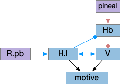
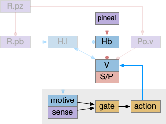
Lateral hypothalamus and value neurotransmitters as a wake hub. Hb (habenula), H.l (lateral hypothalamus), pineal (pineal gland), R.pb (parabrachial nucleus), V (wake-promoting neuropeptides)
The above diagram shows two complementary roles for H.l. First, H.l can suppress Hb.l circadian sleep-promoting path with H.l orexin projections to Hb that inhibit anaesthesia [Zhou et al 2023] and promote aggressive arousal [Flanigan et al 2020].
The positive feedback loop from H.l to Hb (habenula) to V and back to H.l sustains wake. The gain for positive feedback could vary in circadian cycles. A high gain in the morning could produce full wake even with little activity. A low gain at night would make it harder to sustain wake.
Misc notes: Sleep preparation is a distinct, complicated pre-sleep behavior. One trigger seems to be from F.pl (pre limbic frontal cortex) SST neurons to H.l [Tossell et al 2023]. Astrocytes also seem to be involved with H.l wake, are active when waking and promoting wakefulness [Cai et al 2022]. H.pv is also sleep promoting and if it’s knocked out, significant daytime sleep increases, particularly in the morning [Chen et al 2021]. In contrast, the posterior hypothalamus has astrocytes that increase sleep at night [Pelluru et al 2016].
Vta sleep/wake glutamate and GABA
For the moment, let’s ignore Vta (ventral tegmental area) dopamine. Vta includes glutamate and GABA neurons that derive from r1 (hindbrain rhombomere 1) [Lahti et al 2016] that enhance wake with glutamate [Yu et al 2019] and enhance sleep with GABA [Chowdury et al 2019]. The tail of Vta, RMTg (rostromedial tegmental are in r2 / r3) is essential for NREM sleep [Yang et al 2018].
Vta glutamate, GABA, and DA all control sleep using H.l and S.msh (medial shell of the ventral stratum aka nucleus accumbens). As noted above, H.l is a coordinator of sleep and wake with not only orexin but also GABA and glutamate. Inhibiting Vta GABA bypasses sleep homeostasis, producing a mania state during circadian wake times [Yu et al 2021], [Yu et al 2022].
Vta glutamate stimulation promotes continuous wake independent of DA [Yu et al 2019], via projections to H.l, S.sh, and P.v (ventral pallidum), particularly the NOS1 cells.
Vta.g (GABA neurons of Vta) stimulation encourages NREM sleep. If Vta.g are inhibited, the animal remains 100% awake for hours, during the normal wake period, not the normal sleep period [Yu et al 2022]. The Hb.l projection to Vta.g is required for the anesthetic propofol to work [Gelegen et al 2018].
Interestingly, a specific SST (somatostatin) subset of Vta GABA retains a future sleep requirement from social defeat, where social defeat produces extra sleep. When the rodent loses a conflict, these Vta SST neurons have elevated calcium for up to five hours, and then the animal sleeps, these neurons fire to H.l, extending sleep duration [Yu et al 2019]. Speculating here, this multi-hour memory suggests a possible astrocyte involvement.
Returning the dopamine. Vta dopamine produces wake, while Vta dopamine inhibition produces sleep with nesting behavior [Eban-Rothschild et al 2016], as opposed to immediate collapse like narcolepsy. Low dopamine in a behaviorist experiment produces long decays, difficulty in locomotion and sleepiness [Nicola 2007]. However, other studies argue that dopamine itself is not wake promoting [Takata et al 2018].
Habenula
As mentioned in a previous post, Hb (habenula) is a sleep-promoting area as a motor-inhibiting area driven by the pineal gland and extending melatonin’s role [Hikosaka 2012]. This sleep promoting area is in a positive feedback loop with the wake-promoting neurotransmitters and peptides.
Pineal gland through habenula as promoting sleep by suppressing motivation and motor action. Hb (habenula), V (wake-promoting neurotransmitters and peptides).
This above diagram is a different perspective on the prior H.l diagram, where I’ve merged H.l into V and made the motor and motivation suppression explicit. Here the wake-promoting neurotransmitters gate motivation and motor, extending the role of melatonin, which suppresses action.
Active actions promote wake and suppress sleep, like the R.rs wake efferent copies [Teng et al 2022], by stimulating the value neurons. In turn, the value neurons suppress the habenula such as Vta to Hb.l [Webster et al 2021] and serotonin inhibiting Hb.l [Tchenio 2016], H.l orexin and GABA also inhibit Hb.l [Flanigan 2020], [Gazea 2021]. As mentioned above, these positive feedback loops sustain wake despite sleep pressure.
Summary circuit
Putting these components of the sleep/wake circuit together produces something like the following, where I’ve emphasized the habenula to show how that subsystem fit into the whole circuit.
Sleep/wake circuit emphasizing the pineal, melatonin habenula path. Hb (habenula), H.l (lateral hypothalamus), S/P (basal ganglia), Po.v (ventrolateral and median preoptic areas), R.pb (parabrachial nucleus), R.pz (parafacial zone).
As before, the circadian sleep drive from the pineal gland drives the habenula, which inhibits wake neurotransmitters, which inhibits motivation and action using the basal ganglia as a gate. Ongoing action sustains wake against habenula-driven sleep pressure.
The full diagram includes the wake-ignition circuit from R.pb, the sleep-sustain circuit in Po.v (Po.vl and Po.mn), and the wake-sustain circuit in H.l and V. As a reminder, this model is highly simplified and really only serves as a skeleton to organize the various brainstem sleep systems.
Cortical slow wave sleep
Although I’m trying to avoid the cortex as long as possible, studies use cortical slow waves as a sleep marker, so it’s inescapable. Cortical slow waves are globally synchronized neuron firing between around 0.5Hz to 4Hz. The slow wave firing has no information content, but the oscillations may help clear the cortex of metabolic toxins.
During wake, neurons expend to fill the intercellular space because of the neuron’s ion gradients. Filling the intercellular space means the CSF (cerebral-spinal fluid) can’t clear metabolic toxins [Xie et al 2013]. Slow wave sleep shrinks the neurons allowing fluid to fill the intercellular space, and the oscillations may even help with fluid circulation [Fultz et al 2019].
Cortical sleep appears strongly coupled to astrocytes. Astrocyte calcium precedes slow waves in the cortex [Poskanzer and Yuste 2016]. Astrocytes may even organized SWS waves across the cortex, using electrical gap junctions to connect to astrocyte neighbors [Vaidyanathan et al 2021].
Wake signals driving cortical wake. C (cortex), H.l (lateral hypothalamus), Hb (habenula), P.bf (basal forebrain), V (wake neuropeptides)
The above diagram shows a simplified cortical wake circuit, although the cortex is also affected by wake neurotransmitters norepinephrine, serotonin and dopamine. In this model the cortex is mostly an appendage of the brainstem sleep circuit, waking when the brainstem wakes.
P.bf (basal forebrain) is a set of GABA and ACh nuclei that activate the cortex, hippocampus, and olfactory bulb. Although P.bf is identified by its ACh neurons, the GABA projections seem to be more important for wake.
Notes: Local cortical sleep pressure is signaled with GABA [Alfonsa et al 2023]. Parts of the cortex can sleep independently [Krueger et al 2013].
Ppt and P.ldt ACh and wake
Although I’ve lumped Ppt (pedunculopontine nucleus) and P.ldt (laterodorsal tegmental nucleus) with the “V” wake promoting areas, they deserve a special mention because of their connection and similarity with P.bf. Ppt and P.ldt are ACh ganglia near the isthmus midbrain-hindbrain boundary. Ppt is part of the MLR, showing the tight connection between locomotion and wake. Ppt feeds into P.bf, the striatum, and other locomotive regions like H.stn.
Interestingly, all Ppt neurons self-generate gamma oscillations through intrinsic channels [Garcia-Rill et al 2015]. So it could be an ignition source of gamma activation in the striatum and cortex.
Yu X, Li W, Ma Y, Tossell K, Harris JJ, Harding EC, Ba W, Miracca G, Wang D, Li L, Guo J, Chen M, Li Y, Yustos R, Vyssotski AL, Burdakov D, Yang Q, Dong H, Franks NP, Wisden W. GABA and glutamate neurons in the VTA regulate sleep and wakefulness. Nat Neurosci. 2019 Jan;22(1):106-119.
As mentioned in the previous post, sleep is often divided into circadian sleep and homeostatic sleep, although this model is an oversimplification in part because of the metabolic cycles [Borbély et al 2016]. Despite the caveats, I think starting from circadian circuits is a good start.
Also as mentioned previously, circadian cycles may have started as an oxidation-reduction cycle to project from oxygen’s toxicity after the Great Oxidation Event [Edgar et al 2012]. One of the early solutions is melatonin, a powerful natural antioxidant [Tosches et al 2014].
Melatonin
Melatonin exists in almost all animals except sponges. Along with its antioxidant properties, it signals for the zooplankton diel vertical migration, swimming toward the light at dusk and sinking at night [Tosches et al 2014].
System for melatonin-controlled zooplankton vertical migration. ACh (acetylcholine).
In the migrating zooplankton, melatonin triggers ACh (acetylcholine) neurons, which rhythmically spike and these spikes disrupt the cilia, disorganizing them and allowing the plankton to sink.
Reptile and mammal complications
As a complication to understanding the vertebrate circuits, both reptiles and mammals have sleep requirements at odds with aquatic vertebrates. Because land temperatures change more than water temperatures, and reptiles are cold-blooded, their sleep and wake is necessarily strongly tied to temperature as well as the common light/dark connection [Rial et al 2022]. So, sleep and temperature are highly correlated, which makes the Poa (preoptic area) combination of temperature and sleep functions more reasonable than a seemingly random combination.
Mammals have the additional complication of the evolutionary nocturnal bottleneck [Rial et al 2022], meaning the simple heuristic of nighttime melatonin for sleep isn’t sufficient. The pineal melatonin is still at night for nocturnal animals [cite], and the light signaling needs to flip. Although diurnal mammals are no longer nocturnal, their clock circuitry retains the heritage of a nocturnal flip.
As a specific example, all non-mammalian vertebrates use the pineal gland as a circadian oscillator, not H.scn (subthalamic nucleus) [Vatine et al 2011].
Pineal gland and habenula
The pineal gland in the midbrain is the vertebrate’s main source of melatonin. Evolutionarily, the pineal gland is derived from photosensitive cells that directly convert light and dark into melatonin. In non-mammals, the pineal gland is still photosensitive. In the zebrafish, the pineal photoreceptor is still effect and entrains circadian cycles [Vatine et al 2011], and an analogous region in the non-vertebrate chordate Amphioxus provides a similar function, showing the pineal gland’s conserved function in vertebrates [Lacalli 2022].
Hb.m and Hb.l (medial and lateral habenula) derive from the pineal complex, and may have originally been effectors of the pineal gland, serving a nervous function analogous to melatonin [Hikosaka 2010]. Hb.l in particular is well-suited to control neurotransmitters associated with wake, such as dopamine from Vta (ventral tegmental area), serotonin from V.dr and V.mr (dorsal and medial raphe), and norepinephrine from V.lc (locus coeruleus). Note that melatoninAs explored in essay 20, Hb.m is involved in primitive phototaxis and chemotaxis and is well-placed to inhibit those actions during sleep.
Habenula control of sleep by gating motive from action. Hb (habenula), pineal (pineal gland).
In the above diagram, a primitive habenula function is to suppress sensation, motivation and action for sleep by suppression wake-supporting neurotransmitters. Although the diagram illustrates the habenula as disconnecting motive from action, it could also disconnect sense from action, as in phototaxis or chemotaxis in Hb.m.
Hb.m includes an internal entrainable circadian clock, unlike Hb.l. The Hb.m clock is necessary for ultradian foraging. The foraging ultradian is around four hours, generally on waking. Both dopamine and NE (norepinephrine) are elevated [Wang et al 2023] and reciprocally the circadian clock is set by dopamine and NE [Salaberry and Mendoza 2022].
Some misc notes: Hb.l is required for some anesthesia (propofol) and stimulating Hb.l strongly induces NREM, and suppresses motor [Gelengen et al 2018]. Hb.l stimulus produces NREM [Goldstein 1983]. Hb.l is more active mid and late day and early night [Aizawa et al 2013] (possibly producing morning ultradian activity). Hb.l manipulation produces wake fragmentation in the wake period and sleep fragmentation in the sleep period via orexin in H.l [Gelengen et al 2018].
Cell clocks
As mentioned in the introduction, the oxidation-reduction protection may have led to the development of cellular clocks. Essentially all cells have circadian cycle in protein expression, including metabolic and detoxification cells in the liver, heart, kidneys and digestion [Dibner et al 2010], even including gut bioflora. The clocks are synchronized by multiple signals, including feeding patterns, but most studied by light.
For example, dopamine is under clock control and is modulated by melatonin [Ashton and Jagannath 2020]. In S.v (ventrial striatum aka nucleus accumbens) dopamine is at a daily low at night. DAT (dopamine transporter), affected by cocaine, is regulated by clock genes [Alsonso et al 2021], possibly under control of astrocytes. Dopamine is particularly tonically high in early morning before eating with an ultradian cycle of about four hours. Two four-ish hour dopamine cycles are known: the FEO (food entrainable oscillator), which produces pre-feeding activity [Dibner et al 2010], and MASCO (methamphetamine-sensitive circadian oscillatory) [Tataroglu et al 2006], which may be the same system.
The retina itself is under circadian control, modulated by dopamine and D2i (inhibitory Gi-coupled dopamine receptor) [Yujinovsky et al 2006], including in frogs [Cahill and Besharse 1991].
And astrocytes in S.v are under circadian cell clock control [Becker-Krail et al 2022]. Astrocytes are well-placed to manage sleep because they have widespread connections to many synapses and are connected to other astrocytes with gap junctions, allowing for integration over time and space and widespread broadcast signaling.
H.scn circadian entrainment
The circadian system has three distinct components that can either work on their own or work together:
Cell clocks
Light / dark photoreceptors or feeding signals and behavior
Entraining the cell clock to the signal (zeitgeber)
If the eye area of the mollusk sea hare is lesioned, circadian entrainment is eliminated, but because of other photoreceptors, the animal still follows light and dark cycles as long as the light changes. The deficit is only exposed when the lesioned mollusks are placed under continual dark [Vorster et al 2014], [Newcomb et al 2014]. Similarly, in zebrafish many cells are photoreceptive without entraining the cellular clocks.
Mammals use H.scn (suprachiasmatic nucleus) to coordinate circadian cellular clocks. The H.scn name is important, but it’s located above the optic crossing (suprachiasmatic) and developmentally the retina develops from the hypothalamus adjacent to H.scn.
Abstract representation of the mammalian brain highlighting the proximity of the retina and H.scn. arc (H.arc – arcuate nucleus), C (cortex), CB (cerebellum), H.l (lateral hypothalamus), ip (R.ip interpeduncular nucleus), mb (H.mb mammillary bodies), MHB (midbrain-hindbrain boundary), P (pallidum), S (striatum), scn (suprachiasmatic nucleus), sum (supramammillary nucleus), Vta (ventral tegmental area), r1 (rhombomere 1), ZLI (zona-limitans intrathalamica)
The diagram above shows the rough location of the retina development area and H.scn, which both develop from the hypothalamus. A primitive eye with only a few photoreceptors would have been part of the hypothalamus, and like the mollusk the photoreceptor would be near the clock entrainment circuit that became H.scn.
The H.scn clock signal is somewhat indirect, with an interim projection to H.scz to H.dm (dorsomedial hypothalamus) and finally to H.l (lateral hypothalamus) for wake and Po.vl (ventrolateral preoptic area) for sleep. H.scn uses dopamine from Vta as part of its synchronization [Grippo et al 2017].
Ultradian DA – morning foraging
The sleep / wake cycle has an additional boost during normal foraging times such as immediately after waking. In the subjective morning (dark for rodents), wake is encouraged, homeostatic sleep is suppressed, and dopamine levels are higher. After the foraging boost ends, but still in the wake period, tonic dopamine levels drop and the animals take more frequent naps. This hut radian boost of about four hours affects learning and behavior as well as modulating drug abuse [Ruby et al 2013].
Sleep / wake cycle showing morning boost. ZT (zeitgeber time).
Because this ultradian foraging boots wake and suppresses sleep significantly, studies that stimulate or inhibit sleep and wake can specifically affect the ultradian boost without affecting other sleep / wake periods. So it’s very important to look at the hourly effects because the experimental modulation might reduce the foraging boost specifically, but a summary might show a general sleep increase.
H.scn circadian entrainment uses dopamine. DA from either Vta [Grippo et al 2017], [Tang et al 2022] and/or H.sum (supramammillary nucleus) [Luo et al 2018] can entrain food circadian cycles. Note that since the dopamine “A10” area extends beyond the Vta to include H.sum and M.pag.v on opposite ends of the Vta, these studies may be reporting the same area.
The inhibitory neurotransmitter GABA is associated with sleep, and many sleep drugs are GABA stimulants. GABA neurons in Snr (substantia nigra pars reticulata), H.zi (zona incerta), Vta, and Po.vl are all associated with sleep. As mentioned above GABA from mitochondria and in Hydra are used as a sleep promoting neurotransmitter.
While GABA is associated with sleep, other major neurotransmitters like NE, DA, 5HT (serotonin), ACh (acetylcholine) and histamine are associated with wake maintenance of the execution of wakefulness. As discussed in the previous post, ongoing actions need to suppress sleep. NE, DA, and 5HT are all maintain wake while the animal is active and drop when the animal is winding down activity to sleep. Cortical wake requires activity in ACh-rich area in Ppt (pedunculopontine nucleus), P.ldt (laterodorsal tegmental nucleus), and P.bf (basal forebrain).
Produced by H.l, orexin (aka hypocretin) appears to be a wake-maintenance peptide since removal of orexin produces narcolepsy. H.l orexin projects to essentially all of the other wake-maintaining neurotransmitters, including NE, DA, 5HT and ACh. Orexin is slow, waking after tens of seconds, while stimulating V.lc NE is around two seconds [Yamaguchi et al 2018]. On counterargument is that orexin can ramp later in the day [Grady et al 2006], [Mogavero et al 2023], which would suggest that it’s not part of the ultradian foraging system, although it’s also highly tied to foraging. (Suggesting I need to read more articles to see if the contradiction has been resolved.)
Although orexin is the most dramatic of H.l wake, H.l also includes wake and sleep producing GABA and glutamate neurons that may be even more important for wake, independent of the orexin function. Unfortunately, H.l is complex enough that the different functions haven’t been fully pulled apart.
Adenosine is a sleep-promoting molecule derived from the energy molecule ATP, and has extensive receptor throughout the brain, notable in the striatum. Because it’s a product of ATP, it measures local neural activity and possibly sleep need. Its measurement of global brain activity for homeostatic sleep seems more questionable, but adenosine does accumulate throughout the wake period in P.bf [Porkka-Heiskanan et al 2000].
Inflammation peptides like IL-1β are also sleep-promoting [Imeri and Opp 2009]. In addition to their inflammation-related sleep, they seem to be part of normal homeostatic sleep signaling. In Drosophila sleep-need astrocytes produce IL-1β as a signaling peptide [Blum et al 2021]. In zebrafish, sleep deprivation correlates with immune signaling [Williams et al 2007].
Next: ignition and maintenance circuits
After this general discussion on sleep wake, the next post will cover some of the specific sleep and wake circuits, particularly those associated with wake ignition, wake maintenance and sleep maintenance.
Edgar RS, Green EW, Zhao Y, van Ooijen G, Olmedo M, Qin X, Xu Y, Pan M, Valekunja UK, Feeney KA, Maywood ES, Hastings MH, Baliga NS, Merrow M, Millar AJ, Johnson CH, Kyriacou CP, O’Neill JS, Reddy AB. Peroxiredoxins are conserved markers of circadian rhythms. Nature. 2012 May 16;485(7399):459-64.
Rial RV, Canellas F, Akaârir M, Rubiño JA, Barceló P, Martín A, Gamundí A, Nicolau MC. The Birth of the Mammalian Sleep. Biology (Basel). 2022 May 11;11(5):734. doi: 10.3390/biology11050734.
Although the touch sense is a baseline implementation of obstacle avoidance, the essay’s animal should avoid obstacles before bumping into them, but it needs a new, ranged sense. The aquatic vertebrate lateral line senses obstacles, peer, predators, and prey at a distance of around the animal’s length. The lateral line is a basal vertebrate sense, lost in land vertebrates that’s a twin sense to weak electro sensation. Adding a lateral line sense to the toy model will let the simulated animal avoid walls before bumping into them.
Lateral line sense
The lateral line is an aquatic-only primitive vertebrate sense lost in terrestrial animals. Like mouse whisker, inner ear hearing and vestibular sensing, it’s a hair-based object-distance sensor that senses obstacles, peers, predators, and prey with a distance of about the animal’s length. Unlike mouse whiskers but like hearing and vestibular, the lateral line doesn’t touch the obstacle directly, but senses water movement, and from that motion can infer the presence of obstacles. The lateral line sensors are hair-like neuromasts that line the animal’s body along the entire length [Braun and Coombs 2000], [Chagnaud et al 2017].
Illustration of the lateral line sense from the animal (blue) sensing a nearby obstacle (grey wall at right).
Although the diagram above implies that the lateral line sense is a simple distance measurement, the actual sense is a complicated measurement of fluid dynamics and how water flow changes near obstacles. To further complicate the sense, the animal’s own motion changes the water flow. All vertebrates with the lateral line have a cerebellum-like nucleus to cancel out self motion called MON (medial octavolateral nucleus). Electrosensing fish have another cerebellum-like nucleus to cancel its own electric field called DON (dorsal octavolateral nucleus) [Bell et al. 2008]. For this essay I’m completely ignoring all these real complications and will simply use lateral line as a distance measurement.
The LL (lateral line) divides into two disjoint circuits, LL.a (anterior lateral line) covering the head before the ear, and LL.p (posterior lateral line) covering the trunk and tail behind the ear. LL.a connects to the hindbrain at R4 and LL.p connects at R6 [Chagnaud et al 2017]. The lateral line directly innervates the R.rs (reticulospinal) fast escape motor control neurons, including the paired giant Mauthner cells in R4 which trigger fast escape [Mirjany et al 2011].
Although hearing, vestibular, and lateral line use hair sensor sells, they are evolutionary independent [Chagnaud et al 2017]. Because both hearing and LL sense water oscillations, their signals overlap in frequency [Braun and Coombs 2000], with hearing sensing frequencies over 50Hz and LL sensing frequencies below 50Hz.
Lateral line circuits
Because this essay only uses LL for obstacle avoidance, it only implements the direct LL to R.rs connection, inhibiting movement toward an obstacle. The full LL circuit includes input to M.ic (inferior colliculus, hearing) and OT (optic tectum, blindsight) for higher-level navigation. The LL output to Hb.m (medial habenula) exists in the lamprey [Stephenson-Jones et al 2012] although possibly only the electrosensory LL and is used for thigmotaxis (wall hugging).
Lateral line circuit. Non-faded areas are implemented in the essay. CB.mon (medial octavolateral nucleus, cerebellum-like), Hb.m (medial habenula), LL (lateral line), M.ic (inferior colliculus / torus semicircular), MLR (midbrain locomotive region), N.sp (spinal motor neurons), R.rs (reticulospinal motor neurons), r4/r6 (hindbrain rhomobomeres 4 and 6).
To break down the circuit, first consider the fast escape Mauthner cell in r4 (hindbrain rhombomere 4), which connects directly to fast-twitch muscles. For an early vertebrate, a nearby predator could trigger the lateral line, which then triggered the Mauthner cell for a fast escape. The essay’s simulation uses the same circuit, but for obstacle avoidance instead of escape because I haven’t added any predators yet.
Next consider the thigmotaxis circuit using Hb.m (medial habenula) for gradient descent, where LL replaces the phototaxis light/dark gradient in essay 24 or chemotaxis odor gradient in essay 26. The lateral line gradient of water movement as the animal nears an obstacle drives Hb.m to balance wall avoidance with wall hugging. Note that the nAChR (nicotinic receptor) from R.ip (interpeduncular nucleus) to P.ldt (laterodorsal tegmental nucleus) is a goldilocks attract / avoid circuit [Wolfman et al 2018], which is the diagram’s Hb.m to MLR connection. At a distance, the weak glutamate-only R.ip to P.ldt signal is attractive, but nearby the stronger ACh amplification switches to aversion. The “nicotinic” is important here, because nicotine as a drug acts here for both nicotine’s attraction at low doses and its aversion at high doses. (Note: in this case “gradient descent” is the correct term, because the animal has a target distance so either too close or too far forms a signed error.)
Next consider the unmodulated LL to M.ic (inferior colliculus / torus semicircularis) to OT (optic tectum) circuit. Even ignoring optic input, OT has an egocentric map of sensory input, including the lateral line. Since the lateral line neuromasts are distributed across the animal’s body and head, each neuromast can give location-specific information about the distance to the obstacle or threat. The LL data becomes a marker on the OT map, enabling a more sophisticated OT navigation, where all the obstacles are marked on the map.
Finally consider the adaptive filter improvement from CB.mon (medial octavolateral nucleus) as a noise-cancelling system, where the noise is self-generated motion affecting the LL signal. CB.mon uses motor efference copies from MLR and B.rs and LL sensor input to predict the self-motion effect on LL. It sends the self-motion prediction to M.ic allowing M.ic to both filter out the self-motion, but also retain the unfiltered input. After filtering out self-motion, the remaining signal better represents the obstacles or threats. Although this signal could be called a prediction error, I think using filtered signal is a clearer and more useful description because the prediction itself is merely instrumental in producing a useful signal.
Cerebellum-like circuits
CB.mon (medial octavolateral nucleus) in the diagram above is a cerebellum-like system [Bell et al 2008], [Montgomery et al 2012]. Cerebellum-like systems are adaptive filters that remove predictable self-generated signals from self-motion. Because LL measures water motion but swimming also produces water motion, LL will primarily measure the animal’s own motion, unless the animal has paused. Since the self-motion would swamp the signal from obstacle, predators, or prey, CB.mon cleans up the signal by filtering out self-motion.
Social lateral line modulation
Modern fish are far more advanced animals than the simulation and famously swim in schools. The proximity of peer fish adds complexity to the lateral line. The fish needs to allow the peer to come nearer than a predator or even an obstacle.
Since lateral line is old, it’s unsurprising that schooling behavior is peptide-modulated. In zebrafish the neuropeptide pth2 reduces lateral line sensitivity and allows for closer schooling [Anneser et al 2020]. This effect is modulated by fish models of social anxiety, where anxious fish use larger distancing [Anneser et al 2022].
I’ve been ignoring distracting cues in the previous essays for simplification. Since the simulated animal only encountered a single odor at a time, it never needed to select one and ignore the other. In essay 26, I’ll implement a very simple first approximation to ignoring distractors, using the P.bf (basal forebrain) control of the Ob (olfactory bulb) as a switchboard to let the selected odor through and inhibit the ignored distractor.
Simulated animal (triangle) encountering two odor plumes (circles).
In the diagram above, the animal (triangle) is seeking food using the purple odor cue as a gradient direction. When it encounters the distractor odor in blue, it should ignore the distractor, otherwise the two odors will mingle into an incorrect summed gradient and the animal will seek in the wrong direction [Cisek 2022].
Temporal chemotaxis
For essay 26, I’m switching chemotaxis (odor seeking) to use the apical temporal gradient search, using Hb.m (medial habenula) and B.ip (interpeduncular nucleus) like the phototaxis in essay 24. The apical system follows the chimera brain model of [Tosches and Arendt 2013], which suggests that odor senses and actions are distinct systems from bilateral tactile senses. For the essays, the shift is from a bilateral, Braitenberg-like [Braitenberg 1984] system to a modulated random walk like the bacterial tumble-and-run.
Olfactory tumble-and-run system using Hb.m and B.ip for temporal gradient direction, and B.rs for the modulated random walk. B.ip interpeduncular nucleus, B.rs hindbrain reticulospinal motor area, Hb.m medial habenula, Ob olfactory bulb.
The above diagram shows the problem with distractor odors. Because the tumble-and-run system uses a single temporal gradient, it necessarily adds both odors together for its input. The summed input goes to the Hb.m (medial habenula) and B.ip (interpeduncular nucleus) system to modulate the random walk direction.
When the animal crosses into the overlapping distractor odor, it will follow the combined signal, distracted from the original seek target. To avoid distraction, the system can either amplify the current odor A, or inhibit the distractors like odor B.
Analogy with nucleus isthmi
An earlier essay 19 also had an attention / distractor problem, with a different issue of action consistency, and used a zebrafish circuit in P.ni (nucleus isthmi) as a solution. In larval zebrafish P.ni works together with OT (optic tectum) to sustain attention on prey during a hunt [Henriques et al 2019]. P.ni is an ACh (acetylcholine neurotransmitter) and GABA (inhibiting neurotransmitter) system that both amplifies the predicted prey location and inhibits surrounding areas.
Nucleus isthmi circuit as adapted by essay 19. ACh acetylcholine, OT optic tectum, Pni nucleus isthmi.
In the above diagram for the essay 19 circuit, a simultaneous left and right touch would select one action at random and sustain that choice for subsequent movement with the P.ni positive feedback circuit. The outputs are crossed because it’s an avoidance circuit: an obstacle on the left triggers a right turn.
Importantly, the positive feedback is modulatory; it doesn’t trigger an action by itself. At a synapse level, ACh triggers mAChR (ACh metabotropic receptor, Gs stimulatory type) on the sensor axon, amplifying the sensor’s neurotransmitter release. The ACh and mAChR act as the decay timer, because they have a slow time constant on the order of a few seconds. If the sensor doesn’t stimulate the circuit, as when successfully avoiding the obstacle, the attention will decay over a few seconds, resetting the system to its original state.
A similar function applies to Ob and P.bf (basal forebrain), where P.bf acts like P.ni to sustain attention to the selected odor. “Basal forebrain” is a general name for a collection of functionally-related subcortical areas in the ventral (“basal”) forebrain, all pallidal-like (P). The specific P areas for the Ob are P.hdb (horizontal diagonal band) and Po.me (magnocellular preoptic area), but I’ll use P.bf for simplicity.
Olfactory bulb as a switchboard
In this model, Ob acts like a switchboard controlled by P.bf. P.bf selects attended odor paths in Ob, where Ob either passes the odor signal to its destination or inhibits the signal if it’s a distractor. P.bf opens and closes gated circuits in Ob.
Although the architecture of the Ob and P.bf circuit resembles the P.ni circuit, Ob appears to rely more heavily on inhibitory GABA for the gating operation, although ACh is also important [Böhm et al 2020], [de Saint Jan et al 2020], [Nunez-Parra et al 2000]. Since this essay is a first cut, simplified model, I’m using a single signal that represents a gating attention / inhibition signal, and glossing over the ACh vs GABA distinction.
Olfactory bulb switchboard using basal forebrain to gate selected odors. B.ip interpeduncular nucleus, B.rs reticulospinal motor, Hb.m medial habenula, Omt mitral/tufted output cells, Osn olfactory sensor neurons, P.bf basal forebrain.
In the above diagram where the switchboard selects odor A and inhibits odor B, the apical seek circuit receives only odor A’s signal. P.bf gates odors from Osn (olfactory sensory neurons) to Omt (mitral/tufted output cells), which then add to form a single signal for the temporal gradient tumble-and-run seek. For simplicity, I’ve shown the P.bf ACh and GABA signal as a simple gating control.
Once the system detects odor A, P.bf configures the switchboard to pass through A and inhibit other odors, locking out the distractor. Because the selecting signals are modulators, they don’t drive a signal until an odor signal arrives. Like the P.ni circuit, attention will timeout as ACh and its slow mACh receptor decay. When the animal leaves the odor plume, the system resets because the absence of odor A collapses the feedback loop.
Although the essay’s switchboard is an improvement over the naive summation of odor signals, it’s still quite limited. There’s no active selection of a best odor, and the system can’t switch to a better odor cue. Also, since the global give-up circuit isn’t integrated with P.bf, giving up on odor A can’t select odor B. Instead the animal must leave the plume and reset the system.
Slightly more complete Ob switchboard
The Ob is a surprisingly complex system; it’s not just a simple odor system. In addition to the P.bf, Opir (olfactory piriform cortex) also modulates the Ob system, and Ob itself has lateral inhibition between Omt (mitral cell output), which is plastic, learning to discriminate odors itself, as well as modulatory input from the serotonin and noradrenaline system.
In the real Ob, many Osn for the same odor feed into a single Ogl (olfactory glomeruli), which provides input to several Omt, all representing the same odor. Each odor feature has its own Ogl system, several hundred in mammals (two in the essay simulation). Ogl is where the neuropil of the Osn axons meet the Omt dendrites, in a fan-in to fan-out system. Also, each Ogl has many inhibitory Opg (periglomerular inhibitors) with multiple variations, and each Omt has several inhibitory Ogc (olfactory granule cells). The basic fan-in and fan-out structure looks like the following diagram.
To break down the diagram, the core of the switchboard circuit is the Osn to Ogl to Omt to output path; everything else is gating to select or inhibit the signal.
Odor gating happens in two locations: modulating Omt’s input dendrite tree in Ogl by Opg and modulating Omt’s output by Ogc (olfactory granular cell). Because each Omt’s input Ogl is shared for several Omt, the Opg inhibition likely affects many or all Omt for a single Ogl. In contrast, the Ogc inhibition is individual, and the Omt and Ogc circuit creates and manages gamma oscillations, which amplifies and reduces noise from the signal.
Although I’m not planning on touching cortical areas for many essays, the Opir (olfactory piriform cortex) modules the Ob switchboard in a similar circuit as B.pf with some difference. Since the Opir input to the many Ogc and many Ogl is not odor selective [Boyd et al 2015], Ogc must learn the meaning of the Opir input through plasticity.
Global give-up circuit
The essay’s task engagement and give-up circuit currently uses H.l (lateral hypothalamus) and Hb.l (lateral habenula) with V.dr (dorsal raphe serotonin) [Hikosaka 2010], [Chowdhury and Yamanaka 2016]. When a seek fails Hb.l suppresses H.l, H.l ends seek, and the animal moves on [Post et al 2022].
Because the global give-up circuit is entirely disconnected from the olfactory selective attention from the essay, giving up means giving up on all odors, not just the current attended odor.
Simulation
For this essay, I refactored much of the simulation code to clean up ideas from previous essays. A new hindbrain module manages the main locomotion like the zebrafish hindbrain motor area [Dunn et al 2016], which is possibly different from the tetrapod / amniote locomotion in the midbrain. Because the essay animal is currently more primitive than amniotes, this simplification seemed appropriate and makes the code organization more clear.
Olfactory locomotion is now random-walk based following apical tumble-and-run, as opposed to the earlier bilateral path through Vta (ventral tegmental area / posterior tuberculum) and OT (tectum). In zebrafish both paths exist, which I might explore later, but this essay is restricted to the apical temporal gradient search.
The seek mode now slows the animal and adjusts the Levy walk parameters to simulate ARS (area restricted search). As I’ll cover in the problems section, switching to seek mode is still hardcoded.
I split the habenula seek from habenula give-up (Hb.m from Hb.l) and pulled the gradient seek and head direction from B.ip into the habenula seek. Conceptually, the habenula seek code now represents Hb.m and B.ip as a single complex.
Simulated odor seeking with target attention and distractor inhibition.
In the screenshot above, the animal is making a u-turn to return to the food when the odor gradient (blue semicircle) is opposite the head direction (black semicircle). In the upper right, the green box outlined in red represents the attended green odor signal, while the white box outline in blue represents the suppressed blue odor. Despite the Osn naively sensing both blue and green odors because the animal is in the overlap area, only the green odor passes through Omt to the seek system.
The square borders around the odor color represent P.bf modulation. Red is attended (100% pass through), blue is inhibited (10% pass through), and grey is unmodulated (50% pass through).
In the diamond-shaped homunculus, the bright blue triangle represents the u-turn nudge.
As the goal vector shows, the guessed goal direction isn’t very accurate, particularly when the animal is making a turn. Currently, the animal continues to update its guess even in the middle of a turn when the odor data and averages are not appropriate for the current direction.
Essay 24, which investigated temporal gradient navigation, raised the question of head direction and navigation. The essay 24 model followed a zebrafish phototaxis experiment by [Chen and Engert 2014] which created a virtual light spot surrounded by darkness. The phototaxis behavior used Hb.m (medial habenula) and B.ip (interpeduncular nucleus) path using 5HT (serotonin) from V.mr (median raphe) as an average integrator [Cheng et al 2016] to generate the gradient without using head direction. Since B.ip receives head direction input [Petrucco et al 2023], essay 25 explores using head direction with the phototaxis gradient.
In the fruit fly drosophila, head direction and goal direction combine in the fan-shaped body to produce motor commands toward the goal [Matheson et al 2022]. Since the vertebrate B.ip connectivity with head direction resembles the fan-shaped body, this essay will use it as a model.
B.ip connectivity
Head direction from B.dtg (dorsal tegmental nucleus of Gudden) and the photo-gradient input from Hb.m would combine in tabular rows and columns in B.ip, if it resembles the fan-shaped body.
B.ip connectivity following a fan-shaped body model. B.dtg dorsal tegmental nucleus of Gudden, B.ip interpeduncular nucleus, B.rs reticulospinal motor command, Hb.m medial habenula.
Head direction encoding
Head direction is necessarily encoded by neurons. Each neuron in the head direction population has a specific direction, and fires when the animal is heading toward the neuron’s preferred direction.
Head direction encoding. Each neuron (colored box) corresponds to a direction. The neuron in the current direction is active, while other directions are silent.
In general, the heading is encoded is an ensemble of neurons, where several neurons around the actual direction fire at different rates (or possibly delayed phases). In the diagram above, the central direction (blue) has a higher activity while neighboring neurons have smaller values [Petrucco et al 2023].
Drosophila uses a coding for its head direction, where the amplitude of the actual direction neuron is close to one and the neurons at orthogonal directions are zero [Westeinde et al 2022]. This sinusoidal encoding enables neuron-friendly transformations and combinations [Touretzky et al 1993] with advantages over neural rate-encoding or phase encoding, particularly in response speed.
Fan-shaped body: allocentric to egocentric
Fruit fly navigation uses its fly-shaped body to combine an allocentric goal direction with the head direction to create motor commands to turn left or right. Egocentric is self-focused and allocentric is other-focused. Allocentric coordinates are animal-independent like North or toward a distant landmark, which egocentric coordinates are relative to the animal, like forward, right or left.
The fan-shaped body has a tabular shape where each column is a head direction and each row is a goal input [Hulse et al 2021]. The fan-shaped body combines the goal vector and the head direction to create motor commands [Westeinde et al 2022].
The fan-shaped body combines head direction with goal vectors to produce motor commands.
By shifting the head direction and combining the sinusoidal encodings of the goal vector, the motor output is a turn toward left or right. In drosophila, there’s a third motor command for a U-turn when the goal is behind the fly. Each motor command is carried by a specific neuron: PFL2.L (left), PFL2.R (right), and PFL3 (U-turn).
In drosophila, there are 18 distinct head direction columns and up to 9 goal rows. The fan-shaped body is also used for motivation calculations like sleep, despite sleep not fitting into the strict tabular model shown above. To create the strict organization, the fan-shaped body has 400 distinct neuron types [Hulse et al 2021].
Constructing goal vectors
In the phototaxis situation as in essay 24 or [Chen and Engert 2014] the goal vector is constructed from the gradient as the animal enters darkness from light and the head direction at that moment.
Captured goal vector (red) when the animal crosses into darkness.
As the diagram above suggests, the stored vector isn’t the true direction from light to dark, but only the sample along the animal’s path. The gradient value is then stored in the goal direction cells.
Storing the goal vector requires gating based on head direction. In zebrafish, serotonin accumulators can be gated by actions and used as a short term memory (5s – 20s) [Kawashima et al 2016]. For the essay, head dir gates serotonin accumulation as a replacement for the action gating.
Storing gradient into the goal vector based on the current goal. The red direction (south-east) gates its associated serotonin accumulator.
Since V.mr (median raphe) neurons produce consistent tonic oscillations, they are ideal for reading the accumulated value. No additional circuitry for the read is necessary.
Essay simulation
Because the essay model is a functional level, not a circuit level, it can use a directional vector encoding: a pair of floating-point numbers for direction and gradient for strength.
The simulation also calculated two averages: a short-term average for the goal vector gradient and a long-term average for phototaxis gradient motivation. The goal vector average needs to be shorter to avoid bleed-over from a previous direction.
Screenshot of animal crossing into darkness.
The above screenshot shows the animal’s state when it crosses into darkness. The long-timescale motivational gradient (“gr/grad”) is negative, driving the animal to avoid darkness. The short directional gradient (“sa”) is near zero, avoiding update of the stored goal vector. (Note: gradients are 0.5-centered for graphing consistency.)
The homunculus diamond in the upper right shows the current head direction (black semicircle pointing north-east) and the avoidance goal vector (orange semi-circle pointing east). Since the animal is heading toward the avoidance direction, it has a U-turn motor command (orange triangle at top). In addition, since the goal vector and head direction are near a right angle, right turns are inhibited (red at lower right). Because locomotion remains exploratory and stochastic, inhibits reduce turn probability but don’t force turns.
Discussion
This essay’s model is more speculative even compared to other essays, because I haven’t found any papers reporting in B.ip head direction behavior other than the base existence of head direction afferents [Petrucco et al 2023]. In particular, the drosophila fan-shaped body is not homologous to B.ip because the pre-vertebrate animal amphioxus lacks either structure. Nevertheless, it’s interesting that a goal gradient vector circuit is at least possible and relatively simple.
Specifically, the goal vector provides an evolutionary step toward hippocampal (E.hc) object vector cells and grid cells, because those are relatively small enhancements over the goal vector. Without a Bi.ip goal vector system as an intermediary step, hippocampal navigation is too big of an evolutionary step with too many concurrent requirements to be likely.
Note that the hippocampal system is strongly connected with the Hb, B.ip, V.mr, B.dtg system from this essay. E.hc (hippocampus), P.ms (medial septum), Hb (habenula), B.ip (interpeuncular nucleus), V.mr (median raphe), B.dtg (head direction) form a strong connected system together with H.sum (supramammilary/ retromammilary nucleus).
Although the previous essays have focused on bilateral locomotion in the style of Braitenberg machines, the chimaera brain hypothesis [Tosches and Adrendt 2013] suggests a distinct apical form of locomotion. The chimaera brain hypothesis suggests that bilaterian brains are the merger of an apical nervous system from the ancestral zooplankton larva state and a blastoporal (bilateral) nervous system from paired muscles along the spinal cord. The apical area contains unpaired light and chemical sensors and the blastoporal area contains bilateral, topographic somatic sense like touch. Apical navigation require a temporal gradient, calculated by sequential sampling because the apical senses are non-directional.
Apical unpaired light sensors and bilateral paired touch sensors in the chimaera brain.
The essays’ simulation scenario is a temporal phototaxis based on the real-time place preference (RTPP) experiments of [Chen et al 2014], where a zebrafish stayed inside a virtual light circle, avoiding surrounding dark area. Temporal phototaxis is gradient-based locomotion, heading toward light and away from darkness by comparing light samples at different times.
Chimaera locomotor
The vertebrate paired eye and paired olfaction are late vertebrate developments. The pre-vertebrate animal amphioxus has only a single frontal eye. Since pre-paired sense animals needed to navigate toward opportunities and away from threats, it’s conceivable that apical random-walk navigation developed before visual navigation.
Dual navigation system based on both temporal gradient-based random walks and bilateral spatial gradient navigation.
Apical gradient
The apical area contains undirected light and chemical sensors. The apical area is based on the zooplankton larval state as shown below, while the bilateral area is based on bilateral worm-like adults structured like the spinal cord of paired muscles and neurons.
Apical zooplankton larva. Note the single sensor on top.
Apical navigation requires following a temporal gradient, calculated by sequential sampling. While bilateral areas can compare left and right senses to calculate a spatial gradient, the single apical sense is restricted to a temporal gradient.
Bacteria tumble and run
Even simple bacteria can follow gradients using a directed random walk strategy called tumble and run [Segall et al 1986]. The bacteria’s flagella have two modes: tumble, turning without moving, and run, moving forward without turning. By alternating tumble with run, the bacteria can search with a random walk. By extending the run phase when the gradient improves, the bacteria can move toward the target.
Importantly, a temporal gradient calculation needs some sort of memory, accumulator or integrator to compare the current value to recent values. In bacteria, tis accumulator is an internal chemical quantity.
Different turning behavior depending on the gradient. Sharp turns moving into darkness and straight movement into light.
Apical and bilateral sensors
Amphioxus is a pre-vertebrate chordate that’s studied to understand vertebrate evolution. Amphioxus does not have a paired eye, instead it has a single frontal eye that amphioxus uses to orient vertically, and also a pineal-like photoreceptor [Lacalli 2020].
The pineal region is near the vertebrate habenula. Amphioxus does not have a habenula, but it does have a nearby motor control neuron, LPN3, with similar genetic markers [Bozzo et al 2023]. Modern vertebrate medial habenula receives undirected light input from the retina, but it seems plausible that an early habenula used the pineal photosensor because both are part of the same epithalamus complex, and only later connected to the newly-paired retina when it developed.
Multiple apical regions
Before diving into the vertebrate areas for apical locomotion, I need to explain why widespread areas can all be apical, including midbrain and hindbrain areas. Vertebrates had two rounds of whole genome duplication [Dehal and Moore 2005], which gives an easy evolutionary opportunity for four apical areas from the genome duplication, in addition to other possible duplications. The xenobot experiments [Blackiston et al 2023] shows that biology can mix multiple copies like the split apical area into a coherent animal: development can be flexible.
Functional vertebrate brain model showing apical areas as marked by fgf8 development transcription factor.
The diagram above is a functional representation of the vertebrate brain with possible apical areas highlighted in blue. Fgf8 is a development growth factor associated with the apical area [Marlow et al 2014]. The four (or five) possible apical area are as follows:
Prefrontal cortex and olfactory bulb.
The caudal isthmus area (r1) at the midbrain-hindbrain boundary, including cerebellum (CB), interpeduncular nucleus (M.ip), midbrain locomotor (MLR, M.ppt, M.ldt), head direction (B.dtg), parabrachial (B.pb), and part of the substantia nigra (Snr).
Habenula (Hb) and pre-thalamic eminence area (P.em) near ZLI. Also, between the septum-diagonal band (P.msdb) and preoptic area (Poa) near the optic (retina) region (R).
The mammilary and supramammilary area of the hypothalamus (H.mb and H.sum).
I’ve listed four regions instead of the five blue areas because the Hb-P.em-P.msdb area are physical closer than the diagram suggests, are split more by the alar/basal division than distance, and is complicated by distortions from the paired optic region.
This essay uses functions from the r1 isthmus area (M.ip and MLR.α / M.ldt), and from the habenula / retinal area (Hb.m, P.em, pineal, and retina). The logic behind their connectivity is a function split-and-pull like taffy or continental drift from a single pre-duplication area, for example, the isthmus apical area duplicating from an original pre-hypothalamus / retinal apical area.
As a note, the supramammilary area (H.sum) is highly connected with the area in this essay, but I’m postponing exploring its functionality for now.
Vertebrate apical and bilateral locomotor
Previously the essays used the bilateral locomotor path, going through the Vta (posterior tuberculum), tectum (OT), and midbrain locomotive region (MLR). The apical path runs through the medial habenula (Hb.m) and the interpeduncular nucleus (M.ip) before reaching the motor neurons.
Bilateral and apical locomotive paths. B.ll lateral line, B.rs reticulospinal motor neurons, B.ss somatosensory, Hb.m medial habenula, M.ip interpeduncular nucleus, MLR midbrain locomotive region, OT optic tectum, V.mr median raphe, 5HT serotonin.
The lamprey’s Hb.m supports locomotion for light, odor, and the lateral line [Stephenson-Jones et al 2012]. The lateral line is an aquatic sense for water flow, which allows fish to sense nearby objects. Although the exact functional division of phototaxis isn’t known, Hb.m, M.ip and the serotonin raphe nuclei (V.mr – 5HT) are all required [Cheng et al 2016].
For the essay’s simulation, I’m asking the integration and running average to the V.mr and 5HT, but this is something of a guess, because phototaxis integration hasn’t been measured. In the essay’s model, M.ip translates the light data and 5HT average into a gradient and into action.
Phototaxis actions
When zebrafish enter darkness from light, they immediately produce a large turn (O-bend) and start an area restricted search (ARS) [Fernandes et al 2012]. Later turns are smaller [Chen and Engert 2014].
For the essay, phototaxis gradient modifies the standard random walk. When entering darkness, M.ip increases the turn angle. When entering light, M.ip increases forward movement, extending the run.
Essay simulation showing the animal returning to light from darkness. The graph shows the gradient change when crossing the darkness boundary.
The above screenshot shows the animal crossing into darkness and returning to light. In the graph, “light” is the current photosensor value, “avg” is the running average measured by serotonin neurons, and “grad” is the different between the two. A gradient drop triggers high angle turns. A gradient rise triggers straight movement.
As the heat map in the right shows, this simple system produces real-time place avoidance (RTPA) of darkness. Since the system has no learning, there’s no conditioned place aversion (CPA).
Prethalamic eminence
The full phototaxis circuit in vertebrates is a bit more complicated because light input does through an intermediate area called the pre-thalamic eminence (P.em), which is between the habenula, hypothalamus and thalamus, and it one of the apical areas. Although P.em is not cortical, it provides neurons necessary for cortical development (Cajal-Retzius neurons for L1 patterning) [Marin-Padilla 2015] and neurons for habenula input, the habenula-projecting pallidum (P.hb) [Stephenson-Jones 2016].
Apical navigation paths, simplified in lamprey and extended in mammals. E.hc hippocampus, Hb.m medial habenula, M.ip interpeduncular nucleus, MLR midbrain locomotive region, Ob olfactory bulb, P.em pre-thalamic eminence, P.hb habenula projecting pallidum, R.gc retina ganglion cells.
Retina input goes through P.em to Hb.m for phototaxis. Interestingly, the main input for Hb.m in mammals is via cells that migrate from P.em and become the posterior septum (P.ps) [Watanabe et al 2018], which receives almost all of its input from the hippocampus (E.hc). If the hippocampus is an odor-processing system, then the olfactory bulb (Ob) to E.hc to Hb.m path is a chemotaxis path matching the retina’s phototaxis path.
Note that the olfactory placed develops from the lens placed and is differentiated by fgf8 [Bailey et al 2006]. So, it’s pleasing that the similar olfactory and hippocampal paths to Hb.m is a are chemotaxis and phototaxis paths split from a common ancestor.
Speculation
Although the lateral and medial habenula are chemically, connectional, and developmentally distinct, their broad similarity is interesting. If the medial habenula supports direct, concrete sensory navigation by gradient descent, perhaps the medial habenula supports more abstract value-based navigation for more abstract goals.
After essay 21 changed the animal’s default movement to a Lévy exploration, it’s immediate to ask whether that random search is a full action, just like a seek turn or an avoid turn. An if exploration is a controlled action, then the model needs to treat exploration as a full action, like approach or avoid.
Exploration as a full locomotive system at the level of approach and avoid.
[Cisek 2020] identifies a vertebrate system for exploration, including the hippocampus (E.hc) and its associated nuclei such as the retromammilary hypothalamus (H.rm aka supramammilary). Essay 22 considers the idea of treating the subthalamic nucleus (H.stn) as part of the exploration circuit.
Subthalamic nucleus
H.stn is a hypothalamic nucleus from the same area as H.rm, which is part of the hippocampal theta circuit, which synchronizes exploration and spatial memory and learning. However, H.stn is part of the basal ganglia and not directly connected with the exploration system.
[Watson et al. 2021] finds a locomotive function of H.stn, where specific stimulation by the parafascicular thalamus (T.pf) to H.stn starts locomotion. If the stimulation is one-sided, the animal moves forward with a wide turn to the contralateral side. T.pf includes efference copies of motor actions from the MLR as well as from other midbrain actions.
Locomotion induced in the H.stn by T.pf stimulation. H.stn sub thalamic nucleus, T.pf parafascicular nucleus, MLR midbrain locomotor region.
For essay 22, let’s consider the H.stn locomotion as exploration. Since H.stn is part of the basal ganglia, the bulk of essay 22 is considering how exploration might fit into the proto-striatum model of essay 18.
Striatal attention and persistence
Since the current essay simulation animal is an early Cambrian proto-vertebrate, it doesn’t have a full basal ganglia. Evolutionarily, the full basal ganglia architecture could not have sprung into being fully formed; it must have developed in smaller step. Following a hypothetical evolutionary path, the essays are only implementing a simplified striatal model, adding features step-by-step. Unfortunately, because there’s no living species with a partial basal ganglia — all vertebrates have the full system — the essay’s steps are pure invention.
The initial striatum of essay 18 was a partial solution to a simulation problem: persistence. When the animal hit a wall head on, activating both touch sensors, it would choose randomly left or right, but because the simulation is real-time not turn-based, at the next tick both sensors remained active and the animal would choose randomly again, jittering at the wall until enough turns of the same direction escaped the barrier.
Proto-striatum for persistence by attention. Action feedback biases the choice to the last option: win-stay. B.rs reticulospinal motor command, Ob olfactory bulb, MLR midbrain locomotor region, Snc substantia nigra pars compacta (posterior tuberculum).
The main sense-to-action path is from the olfactory bulb (O.b) through the substantia nigra (Snc aka posterior tuberculum in zebrafish) to the midbrain locomotor region (MLR) and to the reticulospinal motor command neurons (B.rs), following the tracing and locomotive study of [Derjean et al. 2010] in zebrafish and Vta/Snc control of locomotion in [Ryczko et al. 2017]. The proto-striatum circuit is built around that olfactory-seeking circuit, acting persistent attention.
The proto-striatal model uses an efference copy of the last action from the MLR to bias the choice of the next action via a MLR to T.pf to striatum path. The model biases the choice through removing inhibition of the odor to action path. If the last action as left, the left odor is disinhibited, making it more likely to win.
The striatal system uses disinhibition for noise reasons. [Cohen et al. 2009] studied attention in the visual system and found that attention removed coherent noise by removing inhibition. By removing inhibition, the attended circuit is less affected by the controlling circuit’s noise.
Note: essay 19 considered an alternative solution to the attention issue by following the nucleus isthmi system in zebrafish as studied in [Grubert et al. 2006], where the attention to the win-stay odor used acetylcholine (ACh) amplification to bias the choice.
Striatal columns: approach and avoid
An immediate difficulty with the simple proto-striatal model is the lack of priority. Although left vs right have equal priority, avoiding a predator is more important than seeking a potential food source. Unfortunately, the proto-striatum treats all options equally. As a solution, essay 18 split the striatum into columns, where each column resolves an internal conflict without priority (“within-system”) and the columns are compared separately (“between-systems”), where “within-system” and “between-system” are from [Cisek 2019].
Dual striatum column for approach and avoid, where MLR resolves the final conflict. B.rs reticulospinal command neuron, B.ss somatosensory (touch), MLR midbrain locomotive region, M.pag periaqueductal gray, Ob olfactory bulb, S.ot olfactory tubercle,S.d dorsal striatum.
Subthalamic nucleus and exploration
If we now treat exploration as a distinct action system, then it needs its own control system and column in the proto-striatum. The within-system choice for exploration is the left and right turns for a random walk, and the between-system choices are between the exploration system and the odor-seeking system.
As a possible neural correlate of exploration, consider the sub thalamic nucleus (H.stn). The sub thalamic nucleus is derived from the hypothalamus, specifically from the same area as the retromammilary area (H.rm aka supramammilary), which is highly correlated with hippocamptal theta, locomotion and exploration.
[Watson et al. 2021] finds a locomotive function of H.stn, where specific stimulation by the parafascicular thalamus (T.pf) produces locomotion via the midbrain locomotive region (MLR). T.pf includes efference copies of motor actions from the MLR as well as other midbrain action efference copies. In the proto-striatum model, the feedback from MLR to striatum uses T.pf.
Exploration locomotive path through H.stn. H.stn sub thalamicnucleus, MLR midbrain locomotive region, T.pf parafascicular thalamus.
Seek and explore with dual striatal columns
Suppose the striatum manages both odor seeking (chemotaxis) and default exploration (Lévy walk). The two actions are conflicting with a complex priority system. When a food odor first appears, the animal should seek toward it (priority to seek), but if no food exists the animal should resume exploration (priority to explore). To resolve the between-system conflict, the two strategies need to columns with lateral inhibition to ensure that only one is selected.
Dual striatum columns for seek and explore strategies. B.rs reticulospinal motor command, H.stn sub thalamic nucleus, Ob olfactory bulb, P.ge globus pallidus external, S.d1 direct striatum projection, S.d2 indirect striatum projection, Snc substantia nigra pars compacta, Snr substantia nigra pars reticulata.
Selecting the seek column enables the odor sense to MLR path, seeking the potential food odor. Selecting the explore column enables the H.stn to MLR path, randomly searching for food.
Note: the double inversion in both paths is to reduce neuron noise [Cohen et al. 2009]. Removing inhibition reduces noise, where adding excitation would add noise. In the essay stimulation, this double negation isn’t necessary.
Striatum with dopamine/habenula control
The previous dual column circuit isn’t sufficient for the problem, because it lacks a control signal to switch between exploit (seek) and explore. The striatum dopamine circuit might help this problem by bringing in the foraging implementation from essay 17.
A major problem in essay 17 was the tradeoff between persistence and perseverance in seeking an odor. Persistence ensures that seeking an odor will continue even when the intermittent. Perseverance is a failure mode where the animal never gives up, like a moth to a flame. As a model, consider using dopamine in the striatum as persistence or effort [Salamone et al. 2007], and control of dopamine by the habenula as solving perseverance with a give-up circuit.
Explore and exploit (seek) columns controlled by dopamine. H.l lateral hypothalamus, Hb.l lateral habenula, H.stn sub thalamic nucleus, MLR midbrain locomotive region, Ob olfactory bulb, P.em pre thalamic eminence, P.ge globus pallidus external, S.d1 striatum direct projection, S.d2 striatum indirect projection, Snc substantia nigra pars compacta, Snr substantia nigra pars reticulata.
The striatum uses two opposing dopamine receptors named D1 and D2. D1 is a stimulating modulator though a G.s protein path, and D2 is an inhibiting modulator through a G.i protein path. In the above diagram, high dopamine will activate the seek column via D1 and inhibiting the explore column via D2. Low dopamine inhibits the seek column and enables the explore column. So dopamine becomes an exploit vs explore controller.
In many primitive animals, dopamine is a food signal. In c.elegans the dopamine neuron is a food-detecting sensory neuron. In vertebrates, the hunger and food-seeking areas like the lateral hypothalamus (H.l) strongly influence midbrain dopamine neurons both directly and indirectly. Indirectly, H.l to lateral habenula (Hb.l) causes non-reward aversion [Lazaridis et al. 2019].
For the essay, I’m taking H.l as multiple roles (H.l is a composite area with at least nine sub-areas [Diaz et al. 2023]), both calculating potential reward (odor) via the H.l to Vta/Snc connection, and cost (exhaustion of seek task without success) via the H.l to Hb.l to Vta/Snc connection.
Lazaridis I, Tzortzi O, Weglage M, Märtin A, Xuan Y, Parent M, Johansson Y, Fuzik J, Fürth D, Fenno LE, Ramakrishnan C, Silberberg G, Deisseroth K, Carlén M, Meletis K. A hypothalamus-habenula circuit controls aversion. Mol Psychiatry. 2019 Sep
Although the essays have implemented obstacle avoidance, they haven’t yet explored olfactory avoidance. Olfactory avoidance is distinct from obstacles, not just because obstacles have higher priority, but because the olfactory system is from an entirely different nervous system than the sensorimotor system. In the chimaeral brain theory [Tosches and Arendt 2013], bilaterian brains are composed of an apical nervous system (ANS) focused on chemo senses (olfactory external and hypothalamic internal), and a blastoporal nervous system (BNS) focused on sensorimotor control like obstacle avoidance.
Olfactory path
The paths for olfactory motion compared with obstacle motion shows the value of the chimaeral theory in making sense of the brain. Working backward from the midbrain locomotive region (MLR), the acetylcholine (ACh) MLR nuclei specialize: the pedunculopontine nucleus (M.ppt) supports the sensorimotor BNS, and the laterodorsal tegmental nucleus (M.ldt) supports the chemosensory ANS.
Sensor-locomotion paths: olfactory on top and somatosensory on bottom. B.ll lateral line, B.rs reticulospinal motor command, B.ss somatosensory, Hb.m medial habenula, M.ldt laterodorsal tegmental nucleus, M.ppt pedunculopontine nucleus, Ob.m medial olfactory bulb, OT tectum, R.vis visual input, ,Vta ventral tegmental area.
In the above diagram, food odors and warning odors use distinct paths to the MLR. Food odors from the olfactory bulb (Ob) pass through the ventral tegmental area (Vta – posterior tuberculum in zebrafish) to the MLR [Derjean et al. 2010]. Aversive odors like cadaverine pass through the medial habenula (Hb.m) to the M.ldt portion of the MLR [Stephenson-Jones et al. 2012]. The food and avoidance paths are distinct because hunger and satiety from the hypothalamus modulate the food path, while the avoidance path can pass through unmodulated. These olfactory locomotion paths correspond to the ANS.
Lamprey medial habenula path
All vertebrates share this basic architecture, including the lamprey, one of the most evolutionary-distant vertebrates. [Stephenson-Jones et al. 2012] traced the Hb.m circuit, showing that Hb.m inputs are from the olfactory path, the parapineal (light attraction), and an electron-sensory alarm to the interpeduncular nucleus (M.ip).
Lamprey olfactory warning path through the habenula to the MLR. M.ip interpeduncular nucleus.
The above diagram fills out the olfactory warning path. The interpeduncular nucleus is a key node in the avoidance circuit, and also key to locomotor-induced theta, and one of the two serotonin nodes. Mip has a major output to the serotonin areas: dorsal raphe (V.dr) and medial raphe (V.mr) and to the central grey (M.pag) [Quina et al. 2017] and M.ldt as well as structures associated with hippocampal (E.hc) theta [Lima et al. 2017].
Medial habenula behavior
In larval zebrafish, Hb.m supports olfactory avoidance [Choi et al. 2017], [Jeong et al. 2021], and light seeking [Zhang et al. 2017]. At least one study indicates that it may also affect food seeking [Chen et al. 2019]. The non-Ob input to Hb.m — the posterior septum (P.ps) — produce locomotion when stimulated [Ostu et al. 2018], suggesting that later evolved functionality maintains the original basal function.
In zebrafish, M.ip only projects to serotonin areas (V.dr and V.mr), not to dopamine or MLR areas. The lamprey connectivity suggests that the M.ip to M.ldt connection was lost in fish.
The Hb.m to M.ip connection is affected by nicotine. An interesting property is that low stimulation and high stimulation have opposite effects. Low stimulation uses glutamate connections and is attractive while high stimulation adds ACh and is aversive [Krishnan et al. 2014].
Developmental genetic notes
As an interesting aside, both Hb.m and avoidant layers of OT shared a genetic marker Brn3a (aka pou4f1) [Quina et al. 2009], [Fedtsova et al. 2008]. That marker also appears in the cerebellum’s inferior olive, trigeminal sensory areas, and the amphioxus motor LPN3 neuron [Bozzo et al. 2023].
M.ldt and M.ppt are sibling areas, deriving from the r1 rhombic lip [Machold et al. 2011].
Glutamate and GABA neurons in M.ip, Vta, and M.ldt all derive from r1 basal neurons [Lahti et al. 2016].
Locomotion switchboard
The addition of olfactory avoidance further complicates the switchboard combining the various locomotor streams, especially if the olfactory path uses serotonin as a modulator as opposed to a straight glutamate connection. Although I’ll probably use a fixed priority for essay 20, and as [Cisek 2022] notes, avoidance can be combined additively, at some point the switchboard will need more control, especially when essays add vision and consummatory actions.